Stabilisation of biological samples

a biological sample and stabilisation technology, applied in the field of biological sample stabilisation, can solve the problems of severe alteration of the expression profile of the targeted molecules, compromise of subsequent analysis, and inability to achieve any practicable methods in hospitals, doctor surgeries or diagnostic routine laboratories, so as to reduce the yield of such nucleic acids and reduce the recovery of such stabilized nucleic acids

Pending Publication Date: 2022-11-17
QIAGEN GMBH
View PDF0 Cites 0 Cited by
  • Summary
  • Abstract
  • Description
  • Claims
  • Application Information

AI Technical Summary

Benefits of technology

[0184]The methods and compositions disclosed herein allow for the efficient preservation and isolation of extracellular nucleic acids while reducing possible mixing with nucleic acids, in particular fragmented genomic DNA, which originates from cells comprised in the biological sample and which may enter a biological sample due to cell damage, respectively cell lysis. The methods according to the present invention, as well as the compositions and the disclosed devices (e.g. the collection containers) reduce the degradation of extracellular nucleic acids and also reduce cell lysis and / or release of genomic nucleic acids, in particular fragmented genomic DNA, so that the extracellular nucleic acids contained in the sample do not become contaminated with intracellular nucleic acids, respectively a respective contamination is reduced by the teachings according to the present invention. As discussed above, an intermixing of extracellular nucleic acids and intracellular nucleic acids, in particular fragmented genomic DNA, may reduce the accuracy of any measurement of the amount of extracellular nucleic acids in a biological sample. As discussed above, an important advantage of the present invention is the possibility for essentially simultaneous stabilizing of both the cells contained in the sample (in particular white blood cells or types of white blood cells in case of whole blood, plasma or serum) and the extracellular nucleic acids. This helps to prevent cellular nucleic acids such as genomic DNA from being released into the cell-free portion of the sample, and further diluting the comprised extracellular nucleic acids (and associated biomarkers) of interest, while also maintaining the structural integrity of the extracellular nucleic acids. As discussed herein, contacting the cell-containing biological sample such as whole blood or plasma with the stabilising agent(s) allows the sample to be stored for a period of time prior to isolating the extracellular nucleic acids. More preferably, the cell-containing biological sample, e.g. blood or plasma, may be drawn at one location (e.g., a health care facility), contacted with the stabilising agent(s), and later transported to a different remote location (e.g., a laboratory) for the nucleic acid isolation and testing process. Furthermore, the stabilization technologies described herein allow to stabilize intracellular nucleic acids and in particular transcript levels as was described in detail above. The stabilization of the transcriptome that can be achieved with the methods and composition described herein is a further important advantage. According to one embodiment, formamide is used for stabilizing the transcriptome. As described above and shown in the examples, formamide is highly effective in stabilizing the transcriptome as well as the extracellular nucleic acid population. Furthermore, cells can be isolated from the sample thereby allowing the analysis of specific cell populations such as e.g. tumor cells, e.g. circulating tumor cells in blood samples. The advantages and technical effects were described in detail above and it is referred to the above disclosure.
[0185]Furthermore, the stabilization reagents and methods, as disclosed in herein, provide an advantage over known state-of-the-art stabilization reagents and methods which involve the use of cross-linking reagents, such as formaldehyde, formaldehyde releasers and the like, as the stabilization of samples according to the present invention does not involve the use to such crosslinking reagents. Crosslinking reagents cause inter- or intra-molecular covalent bonds between nucleic acid molecules or between nucleic acids and proteins. This effect can lead to a reduced recovery of such stabilized and partially crosslinked nucleic acids after a purification or extraction from a complex biological sample. As, for example, the concentration of circulating nucleic acids in a whole blood samples is already relatively low, any measure which further reduces the yield of such nucleic acids should be avoided. This may be of particular importance when detecting and analyzing very rare nucleic acid molecules derived from malignant tumors or from a developing fetus in the first trimester of pregnancy. Therefore, no formaldehyde releaser is comprised in the stabilizing composition, respectively is not additionally used for stabilization. Thus, according to one embodiment, no cross-linking agents such as formaldehyde or formaldehyde releasers are comprised in the stabilizing composition, respectively are not additionally used for stabilization. Thus, here the stabilization composition does not comprise a cross-linking agent that induces nucleic acid-nucleic acid, nucleic-acid-protein, in particular protein DNA and / or protein-protein crosslinks. In particular, the stabilization composition does not comprise formaldehyde, formaline, paraformaldehyde or a formaldehyde releaser. Furthermore, as described, the stabilizing composition does preferably not comprise any additives that would induce the lysis of cells, such as e.g. chaotropic salts. Furthermore, according to one embodiment, the stabilization method according to the invention does not involve the use of additives that classify as toxic agents.
[0186]Particularly preferred aspects and embodiments are described again in the following.
[0187]In a first aspect, the present invention is in particular directed to a method for stabilizing a cell-containing biological sample by contacting the sample with at least one carboxylic acid amide, wherein the carboxylic acid amide is selected from primary carboxylic acid amides and secondary carboxylic acid amides. Preferably, the resulting composition comprising the cell-containing biological sample and the at least one carboxylic acid amide comprises the carboxylic acid amide in a concentration of at least 0.25%. According to one embodiment, the stabilization results in a stabilization of intracellular RNA and / or wherein the extracellular nucleic acid population comprised in the cell-containing sample is stabilized. As described herein, the method is not based on cell lysis and wherein the stabilization does not involve the use of a cross-linking agent that induces protein-nucleic acid and / or protein-protein crosslinks and does not involve the use of a formaldehyde releaser.
[0188]According to one embodiment, the carboxylic acid amide which is selected from primary carboxylic acid amides and secondary carboxylic acid amides has the formula 1
[0189]wherein R1 is a hydrogen residue or an alkyl residue, wherein R2 is selected from a hydrogen residue and a hydrocarbon residue with a length of the carbon chain of 1-20 atoms arranged in a linear or branched manner, wherein R3 is a hydrogen residue, and wherein R4 is oxygen.

Problems solved by technology

However, it was found that the preanalytical steps, such as sample handling and sample stabilisation, in particular for new biomolecular targets, have a severe impact on the expression profile and may compromise the subsequent analysis (see for example Hartel et al, 2001, Pahl and Brune, 2002).
Without precaution in the stabilisation of the sample to be analysed, the sample will undergo changes during transport and storage that may severely alter the expression profile of the targeted molecules (see for example Rainen et al, 2002; Baechler et al, 2004).
These methods are not at all practicable techniques for hospitals, doctor surgeries or diagnostic routine laboratories.
The disadvantage of the respective methods is that the stabilisation results in the complete lysis of the cells.
The destruction of the cells is a great disadvantage because any cell sorting or cell enrichment respectively cell analysis becomes impossible.
However, using different stabilisation reagents and accordingly stabilisation tubes for collecting the sample for nucleic acid analysis and cell analysis is tedious.
However, nucleic acid isolation from respectively stabilised samples is very difficult, because the used formaldehyde releaser interferes with the subsequent nucleic acid isolation process.
Therefore, the nucleic acid yield and / or purity is severely reduced compared to the isolation of nucleic acids that were stabilised using stabilization methods that specifically aim at the stabilization and isolation of nucleic acids such as RNA (for example the PAXgene Blood RNA Tubes).
However, obtaining an essentially cell-free fraction of a sample can be problematic and the separation is frequently a tedious and time consuming multi-step process as it is important to use carefully controlled conditions to prevent cell breakage during centrifugation which could contaminate the extracellular nucleic acids with cellular nucleic acids released during breakage.
Furthermore, it is often difficult to remove all cells.
Once cell lysis begins, the lysed cells release additional nucleic acids which become mixed with the extracellular nucleic acids and it becomes increasingly difficult to recover the extracellular nucleic acids for testing.
Further, the amount and recoverability of available extracellular nucleic acids can decrease substantially over time due to degradation.
However, the use of formaldehyde or formaldehyde-releasing substances has drawbacks, as they may compromise the efficacy of extracellular nucleic acid isolation by induction of crosslinks between nucleic acid molecules or between proteins and nucleic acids.

Method used


Image

  • Stabilisation of biological samples
  • Stabilisation of biological samples
  • Stabilisation of biological samples

Examples


example 1

tion Using Formamide

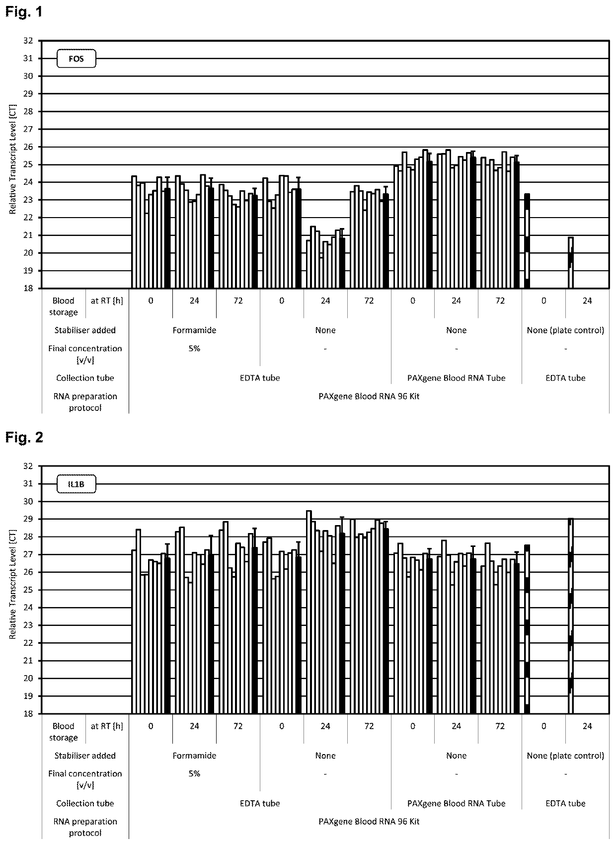
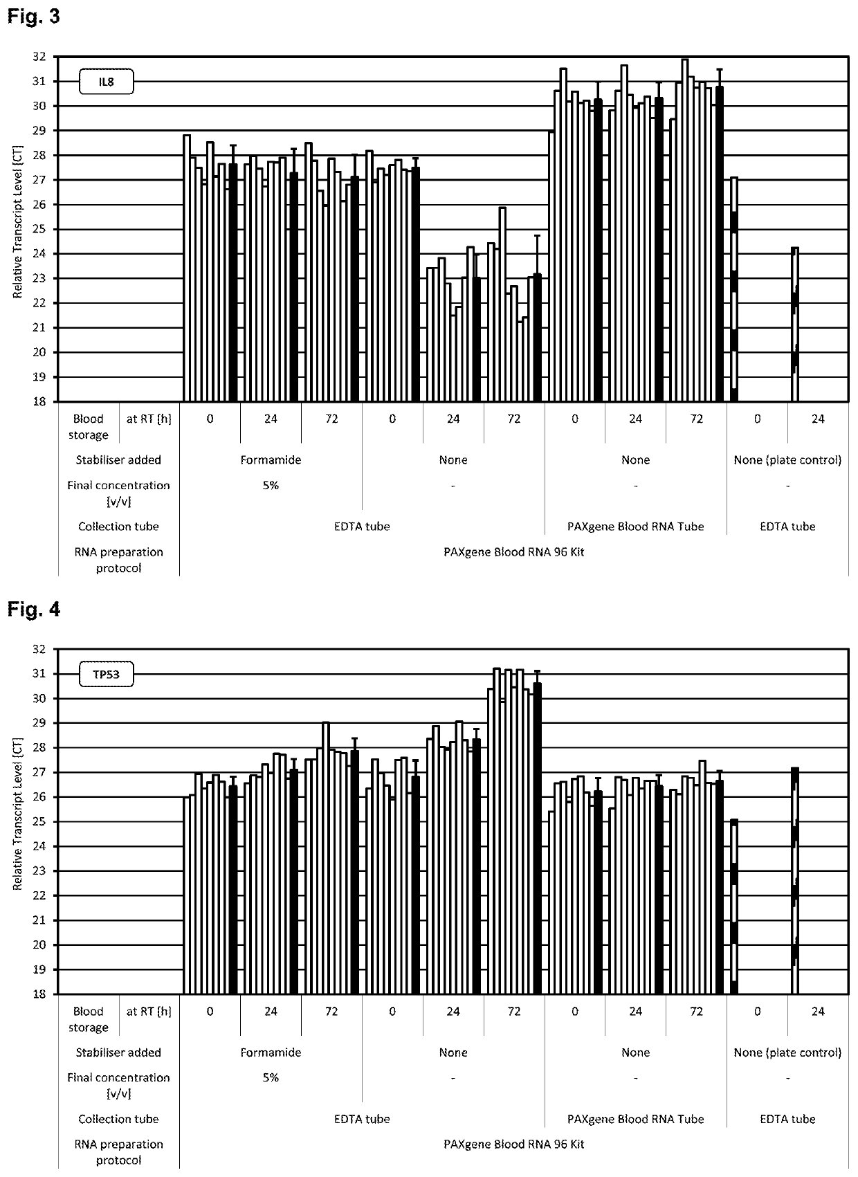
[0280]Blood was collected from multiple donors into replicate EDTA blood tubes (BD, [−] control of sample stabilisation) and PAXgene Blood RNA Tubes (PreAnalytiX) serving as [+] control of sample stabilisation. Immediately after blood collection half of the EDTA blood samples were treated with RNA stabilisation test solution, resulting in blood test samples. In detail, 1 ml stabilisation additive (50% v / v formamide, 5×MOPS buffer, pH5.5) was added to 9 ml EDTA blood. Blood mixing, incubation, transfer of blood sample aliquots to PAXgene Blood RNA Tubes, freezing, storage and RNA preparation was done as described above. RNA was subjected to transcript level analysis as described above in Material and methods. Quantity (RNA yield) and quality (RNA purity, RNA integrity) of RNA was measured by UV spectroscopy and miniaturised capillary gel electrophoresis with RIN calculation (Nanochips on Agilent Bioanalyzer). Transcript levels of all RNA samples were analysed by r...

example 2

tion Using Acetamide

[0286]EDTA blood samples were kept untreated or mixed with 8% w / v acetamide immediately after blood collection. Blood samples collected in PAXgene RNA blood tubes served again as control. Sample handling and RNA isolation was performed from blood samples without and from replicate tubes after incubation for one day at RT as described in material and methods. Transcript levels of all RNA samples were analysed as described in material and methods by real time RT-PCR using monoplex assays of FOS, IL1B, IL8 and TP53, normalized to the amount of template input into the reaction. The results are shown in FIGS. 7 to 10. Resulting CT values reflecting the amount of transcripts were directly compared. Relative transcripts levels unaffected from blood sample incubation at RT were indicated by constant CT values, while gains of transcripts (e.g., by gene induction) were indicated by lower CT and losses of transcripts (e.g., by gene repression) by higher CT values. As can be...

example 3

tion Using N-Methylacetamide

[0287]Samples were prepared and processed as described in example 2, however using 2% (w / v) N-methylacetamide as stabilizer in the composition comprising the blood sample and the stabilising agent. The results are shown in FIGS. 11 to 14. As can be seen, the secondary carboxylic acid amide N-methylacetamide was effective in stabilizing transcript levels for up to three days.

[0288]Examples 1 to 3 show that different primary and secondary carboxylic acid amides are highly effective in stabilizing the gene transcription profile of cells in blood samples.

[0289]II. Stabilization of the Extracellular Nucleic Acid Population in Blood Samples Using Different Primary and Secondary Carboxylic Acid Amides

[0290]Materials and Methods

[0291]Different primary and secondary carboxylic acid amides were tested for their ability to stabilize a cell-containing biological sample, here a whole blood sample, either alone or in combination with a caspase inhibitor. As can be seen...


PUM

PropertyMeasurementUnit
timeaaaaaaaaaa
real time monoplex RT-aaaaaaaaaa

Abstract

The present invention provides methods and composition suitable for stabilizing cell-containing samples such as blood samples. The stabilizers used are primary or secondary carboxylic acid amides.

Description

CROSS REFERENCE TO RELATED APPLICATIONS[0001]The present application is a continuation application of U.S. application Ser. No. 14 / 777,902 filed Sep. 17, 2015, now pending, which is a U.S. national phase application of PCT / EP2014 / 000725 filed Mar. 18, 2014, which claims priority to U.S. Provisional Application No. 61 / 803,107 filed Mar. 18, 2013, EP Application No. 13160896.0 filed Mar. 25, 2013 and EP Application No. 13180130.0 filed Aug. 12, 2013. U.S. application Ser. No. 14 / 777,902 is herein incorporated by reference in its entity.STATEMENT REGARDING SEQUENCE LISTING[0002]The Sequence Listing associated with this application is provided in text format in lieu of a paper copy, and is hereby incorporated by reference into the specification. The name of the text file containing the Sequence Listing is 770025.471C1_SEQUENCE_LISTING.txt. The text file is 25,845 bytes, was created on May 19, 2022, and is being submitted electronically via EFS-Web.[0003]The work leading to this inventio...

Claims


Application Information

Patent Timeline
Patent Type & Authority Applications(United States)
IPC IPC(8): C12Q1/6806C12Q1/6841C12N15/10
CPCC12Q1/6806C12Q1/6841C12N15/10C12N15/1003C12Q2527/125C12Q2527/127C12Q2527/156
Inventor WYRICH, RALFVOSS, THORSTENGÜNTHER, KALLEOELMÜLLER, UWE
Owner QIAGEN GMBH
Why Eureka
Try Eureka