Stabilisation and isolation of extracellular nucleic acids

a technology stabilization, applied in the field of stabilisation and isolation of extracellular nucleic acids, can solve the problems of difficult to remove all cells, difficult to obtain an essentially cell-free fraction of a sample, difficult to obtain all cells, etc., to achieve high efficiency in stabilizing the extracellular nucleic acid population, avoid or at least significantly reduce contamination with genomic dna, and stable cell-containing biological samples

Active Publication Date: 2019-10-10
QIAGEN GMBH
View PDF0 Cites 0 Cited by
  • Summary
  • Abstract
  • Description
  • Claims
  • Application Information

AI Technical Summary

Benefits of technology

[0014]The present invention is based on the finding that certain additives are surprisingly effective in stabilizing cell-containing biological samples comprising extracellular nucleic acids, in particular whole blood samples or samples derived from whole blood such as e.g. blood plasma. It was found that these additives are highly efficient in stabilizing the extracellular nucleic acid population and in particular are capable to avoid or at least significantly reduce contaminations with genomic DNA, in particular fragmented genomic DNA.

Problems solved by technology

However, obtaining an essentially cell-free fraction of a sample can be problematic and the separation is frequently a tedious and time consuming multi-step process as it is important to use carefully controlled conditions to prevent cell breakage during centrifugation which could contaminate the extracellular nucleic acids with cellular nucleic acids released during breakage.
Furthermore, it is often difficult to remove all cells.
Once cell lysis begins, the lysed cells release additional nucleic acids which become mixed with the extracellular nucleic acids and it becomes increasingly difficult to recover the extracellular nucleic acids for testing.
Further, the amount and recoverability of available extracellular nucleic acids can decrease substantially over a period of time due to degradation.
The above discussed problems particularly are an issue, if the sample comprises a high amount of cells as is the case e.g. with whole blood samples.
However, the need to directly separate e.g. the plasma from the blood is a major disadvantage because many facilities wherein the blood is drawn (e.g. a doctor's practice) do not have a centrifuge that would enable the efficient separation of blood plasma.
Furthermore, plasma that is obtained under regular conditions often comprises residual amounts of cells which accordingly, may also become damaged or may die during handling of the sample, thereby releasing intracellular nucleic acids, in particular genomic DNA, as is described above.
These remaining cells also pose a risk that they become damaged during the handling so that their nucleic acid content, particularly genomic (nuclear) DNA and cytoplasmic RNA, would merge with and thereby contaminate respectively dilute the extracellular, circulating nucleic acid fraction.
However, again, such powerful centrifuges are often not available at the facilities wherein the blood is obtained.
This too imposes practical constraints upon the processing of the samples as e.g. the plasma samples must be shipped frozen.
This increases the costs and furthermore, poses a risk that the sample gets compromised in case the cold chain is interrupted.
However, even though EDTA is an efficient anticoagulant, EDTA does not efficiently prevent the dilution respectively contamination of the extracellular nucleic acid population by released intracellular nucleic acids.
Accordingly, EDTA is not capable of sufficiently stabilising the extracellular nucleic acid population in particular because it can not avoid the contamination of the extracellular nucleic acid population with e.g. genomic DNA fragments which are generated after blood draw by cell degradation and cell instability during sample transportation and storage.
However, the use of formaldehyde or formaldehyde-releasing substances has drawbacks, as they may compromise the efficacy of extracellular nucleic acid isolation by induction of crosslinks between nucleic acid molecules or between proteins and nucleic acids.

Method used


Image

  • Stabilisation and isolation of extracellular nucleic acids
  • Stabilisation and isolation of extracellular nucleic acids
  • Stabilisation and isolation of extracellular nucleic acids

Examples


example 1

tion by the Addition of a Caspase-Inhibitor

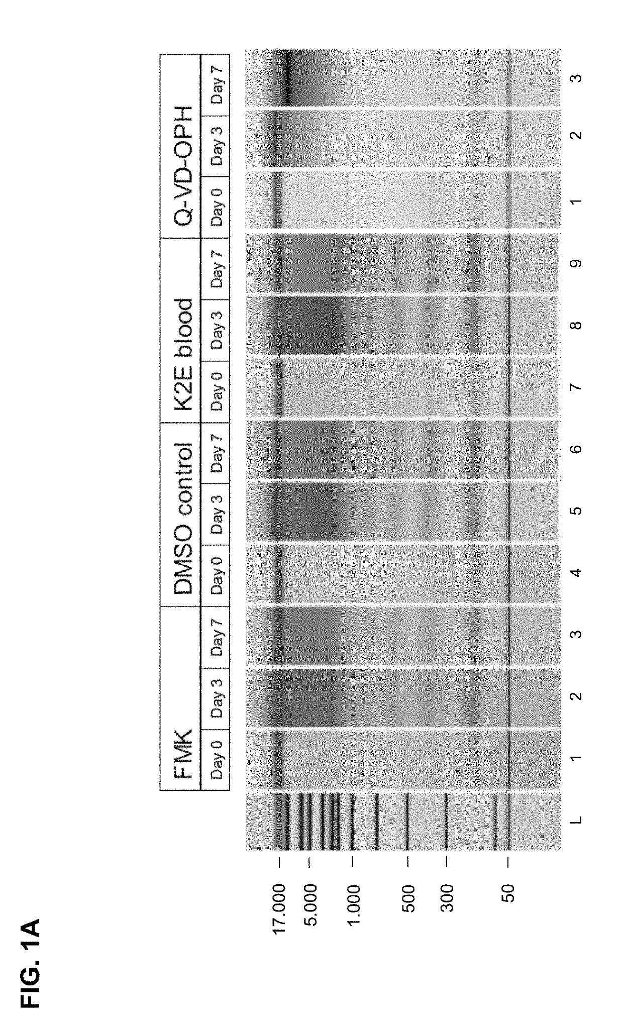
[0230]Two different oligopeptides, Q-VD-OPh and Z-Val-Ala-Asp(OMe)-FMK acting as broad spectrum caspase-inhibitors, were tested:

TABLE 6Tested caspase inhibitorsinhibitormolecularenameweightsolubilitystructureQ-VD-OPH513.4920 mM, add 97 μl DMSO 10 mM, add 194 μl DMSO 5 mM, add 388 μl DMSO Z-Val-Ala- Asp(Ome)- FMK467.4920 mM, add 107 μl DMSO 10 mM, add 214 μl DMSO 5 mM, add 428 μl DMSO

[0231]Each tested caspase inhibitor was added to whole blood samples (20 μM end concentration in 10 ml blood; blood was collected into Vacutainer K2E Tubes; BD). The whole blood sample was processed as described in section I, see 2. (plasma preparation) and 3. (nucleic acid isolation).

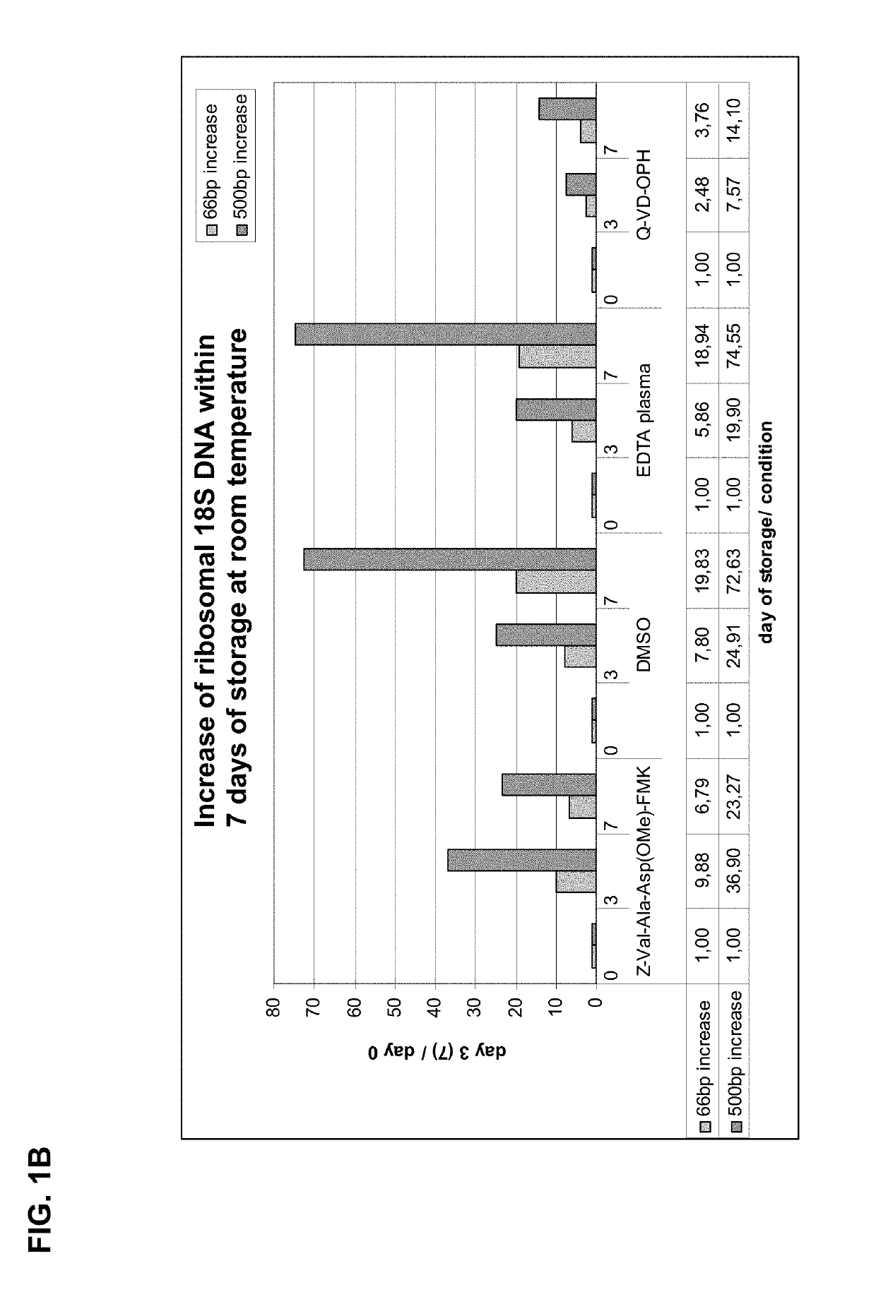
[0232]Results of the Chip Gel Electrophoresis

[0233]The eluted circulating cell-free DNA was separated by size using chip gel electrophoresis (for details on the method see above, I, 4.1). FIG. 1A shows the obtained results. The DMSO control and the K2E blood (not treated according...

example 2

of Lower Concentrations of Caspase-Inhibitor Q-VD-OPh on Blood Stability

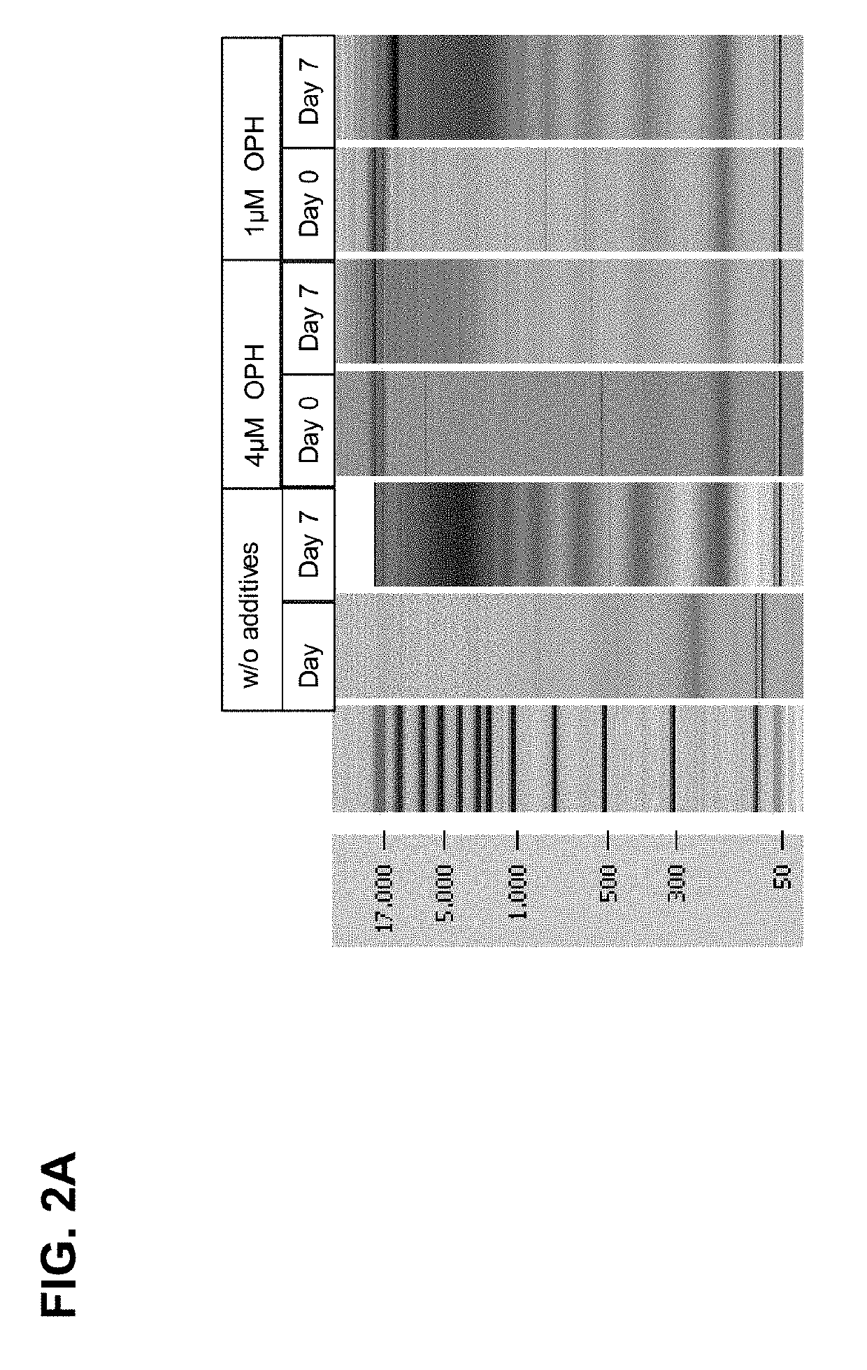
[0241]In this example, lower concentrations of the caspase inhibitor Q-VD-OPh was tested in combination with glucose, wherein the glucose was added as combination partner to support that the blood cells stay alive (by preventing cell damage). 21.4 mM glucose and 4 μM, 1 μM or no Q-VD-OPh were added to 10 ml blood drawn into BD Vacutainer tubes and stored for up to 7 days at room temperature. The whole blood sample was processed as described in section I, see 2. (plasma preparation) and 3. (nucleic acid isolation).

[0242]Results of the Chip Gel Electrophoresis

[0243]The eluted DNA was separated by size using chip gel electrophoresis (for details on the method see above, I, 4.1). FIG. 2A shows that compared to the control samples, wherein no caspase inhibitor was added, already 1 μM caspase inhibitor significantly reduced the genomic DNA release / fragmentation on day 7. The effect is improved if 4 μM caspase inhibito...

example 3

ng Blood Cells by Osmotic Effects

[0247]Surprisingly it was also found by the inventors that blood cells can be stabilized by adding a reagent that acts as a hypertonic medium in whole blood. Generating a hypertonic medium by the addition of, for example, hydroxylated organic compound(s) to whole blood results in a slight release of water from the contained blood cells and results in increased stability by cell shrinking. It is assumed that said cell shrinking stabilises the cells against mechanical forces.

[0248]Dihydroxyacetone (DHA) is an intermediate product of the fructose metabolism and its phosphate form dihydroxyacetone phosphate (DHAP) is part of the glycolysis. DHA was tested as hypertonic agent. Addition of this reagent sensitively forces blood cells to shrink without damaging them. DHA was first dissolved in PBS (purchased from SIGMA-Aldrich Kat. No: D8537) or 3×MOPS (diluted from 1 litre of 10×MOPS: 200 mM MOPS; 50 mM NaAc, 10 mM EDTA; pH 5; assuming that an acid medium a...


PUM

PropertyMeasurementUnit
temperaturesaaaaaaaaaa
temperatureaaaaaaaaaa
elution volumeaaaaaaaaaa

Abstract

The present invention provides methods, compositions and devices for stabilizing the extracellular nucleic acid population in a cell-containing biological sample using an apoptosis inhibitor, preferably a caspase inhibitor, a hypertonic agent and/or a compound according to formula 1 as defined in the claims.

Description

CLAIM OF PRIORITY[0001]This application is a continuation of U.S. application Ser. No. 14 / 347,051, filed on Aug. 7, 2014, now pending, which is a U.S. national phase application of PCT / EP2012 / 068892, filed Sep. 25, 2012, which claims priority to U.S. Application No. 61 / 539,245 filed on Sep. 26, 2011. U.S. application Ser. No. 14 / 347,051 is herein incorporated by reference in its entity.[0002]The work leading to this invention has received funding from the European Community's Seventh Framework Programme (FP7 / 2007-2013) under grant agreement n° 222916.STATEMENT REGARDING SEQUENCE LISTING[0003]The Sequence Listing associated with this application is provided in text format in lieu of a paper copy, and is hereby incorporated by reference into the specification. The name of the text file containing the Sequence Listing is 771025_405C1_SEQUENCE_LISTING.txt. The text file is 23 KB, was created on Nov. 28, 2018, and is being submitted electronically via EFS-Web.FIELD OF THE INVENTION[0004]...

Claims


Application Information

Patent Timeline
Patent Type & Authority Applications(United States)
IPC IPC(8): C12Q1/68C12Q1/6806C12N15/10
CPCC12Q1/68C12N15/1003C12Q1/6806C12Q2527/127
Inventor HORLITZ, MARTINSCHUBERT, ANNABELLESPRENGER-HAUSSELS, MARKUS
Owner QIAGEN GMBH
Why Eureka
Try Eureka