Mandibular occlusal inhibitor

a mandibular occlusal and inhibitor technology, applied in the field of mandibular occlusal inhibitors, can solve the problems of difficult radiographic imaging, difficult diagnosis of spinal injury in patients who are uncooperative, and difficult radiographic imaging,

Inactive Publication Date: 2004-06-01
THE CHILDRENS HOSPITAL OF PHILADELPHIA
View PDF7 Cites 37 Cited by
  • Summary
  • Abstract
  • Description
  • Claims
  • Application Information

AI Technical Summary

Problems solved by technology

Spinal injury may be difficult to diagnose in patients who are uncooperative, have altered mental status, or other distracting injuries.
In such cases, it is difficult to obtain radiographic imagery to make a proper evaluation of the patient.
Obtaining proper cervical spine films, particularly the open-mouth odontoid view (OMV), in the pediatric and adult populations can be a challenging experience even under the best patient circumstances.
Patient-related concerns include inability to cooperate during the procedure, repeat films, excessive radiation exposure, and delays in care.
Although CSI in children is rare, it generally carries a greater degree of morbidity and mortality in the younger pediatric population with associated head trauma.
Failure to adequately diagnose CSI promptly can lead to severe neurologic disability and morbidity.
Two of the more common errors are failure to obtain adequate C-spine roentgenograms and difficulty in x-ray interpretation.
However, obtaining the OMV x-rays can be technically difficult and time consuming for both the patient and the emergency health care provider.
ending on the patient's condition, it may be very difficult to have the patient open their mouth and keep the mouth open while radiography is performed. Pro
blems may be attributed to the patient's age, the patient's mental impairment, unconsciousness, pain or injury that distracts the patient, inability to open the patient's mouth, and difficulties in positioning the patient.

Method used


Image

  • Mandibular occlusal inhibitor
  • Mandibular occlusal inhibitor
  • Mandibular occlusal inhibitor

Examples


Embodiment Construction

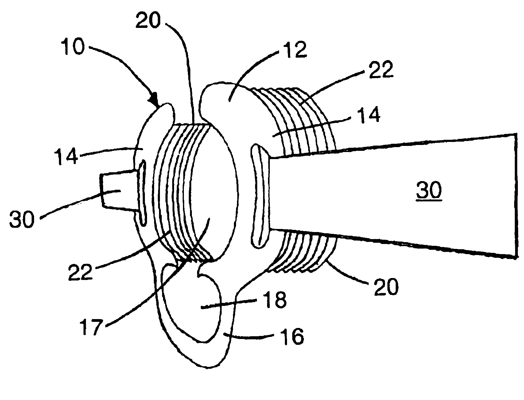
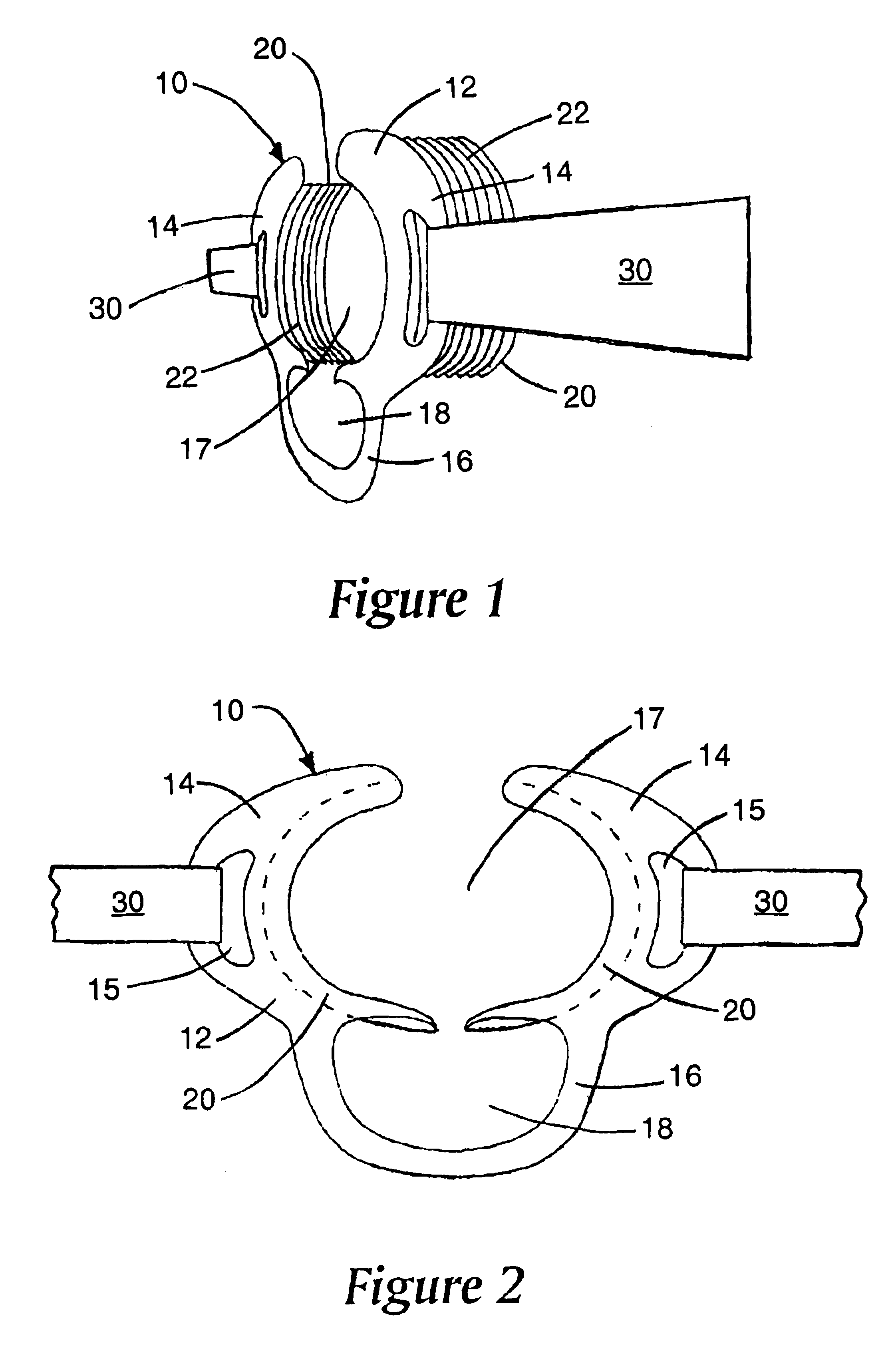
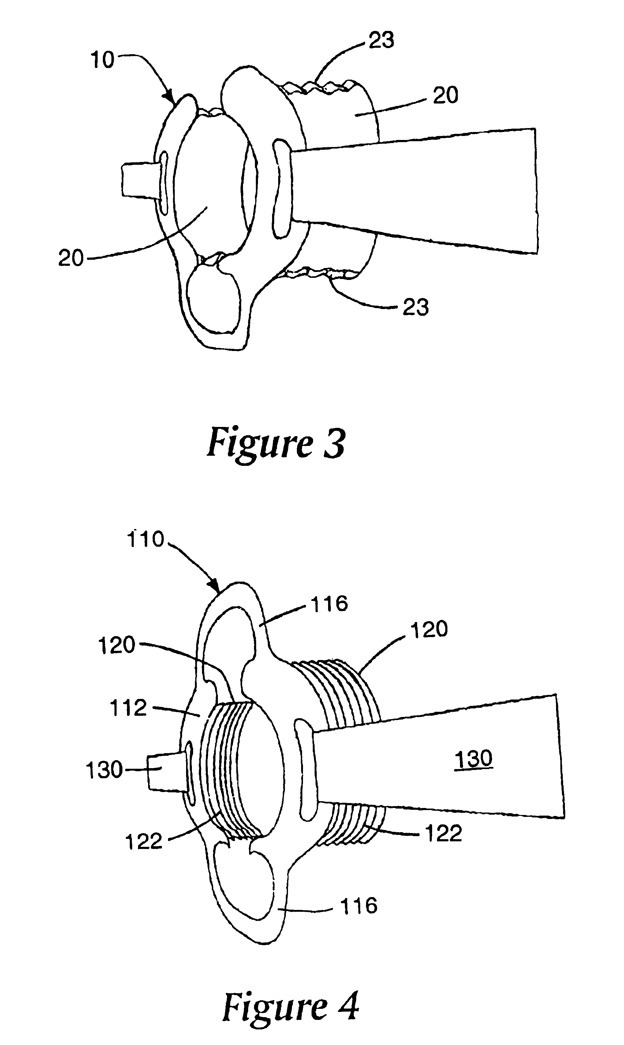
Referring now to FIGS. 1-11 generally, and FIG. 1 in particular, a mouth positioning device in accordance with the present invention is shown and designated generally by reference number 10. Wherever possible, the same reference numbers will be used throughout the drawings and the description to refer to the same or like parts.

FIG. 1 is a perspective view of the mouth positioning device 10. The device 10 is configured to support and maintain a patient's mouth in an open position to permit completion of various medical, dental and surgical procedures. The device 10 may be used in a number of applications, including but not limited to, radiology, endoscopy, dentistry and surgery. In radiology, the device 10 can be used in emergency trauma rooms or in free-standing radiology suites.

Referring now to FIGS. 1-2, the device 10 comprises a body portion 12. The body portion is formed of one or more radiolucent materials that permit passage of radiation or x-rays. In this way, radiographic im...


PUM


Abstract

A device for maintaining a patient's mouth in an open position during a medical procedure is provided. The device includes a cylindrical body configured to be received in the patient's mouth. A flange extending from an edge of the body is configured to engage the face of the patient, and preferably, the device includes a retainer for retaining the body in the patient's mouth.

Description

The present invention relates to a device for holding a patient's mouth in an open position during medical, dental and surgical procedures.DESCRIPTION OF THE RELATED ARTMedical professionals frequently perform procedures that require the patient's mouth to be held in an open position. For example, a patient's mouth must be held open during endoscopic procedures to provide clearance for an endoscope and other instruments. A patient's mouth must also be held in an open or semi-open position when dental bite-wing x-ray images are taken. A similar need exists in trauma evaluation and diagnosis of spinal injuries.Spinal injuries require prompt evaluation by emergency health care providers. Early recognition of possible cervical spine injury (CSI) is of critical importance. Spinal injury may be difficult to diagnose in patients who are uncooperative, have altered mental status, or other distracting injuries. In such cases, it is difficult to obtain radiographic imagery to make a proper ev...

Claims


Application Information

Patent Timeline
Patent Type & Authority Patents(United States)
IPC IPC(8): A61C5/14A61C5/00A61B1/24A61C3/00
CPCA61B1/24A61C5/14A61C5/90
Inventor O'NEILL, KATHLEEN A.
Owner THE CHILDRENS HOSPITAL OF PHILADELPHIA
Why Eureka
Try Eureka