Non-isotopic detection of osteoblastic activity in vivo using modified bisphosphonates

a technology of osteoblastic activity and bisphosphonates, which is applied in the direction of diagnostic recording/measuring, ultrasonic/sonic/infrasonic diagnostics, drug compositions, etc., can solve the problems of radioactive compound use, not providing high-resolution anatomical detail for radioscintigraphic detection, and not being able to directly detect ha in vivo

Inactive Publication Date: 2005-03-22
BETH ISRAEL DEACONESS MEDICAL CENT INC
View PDF7 Cites 53 Cited by
  • Summary
  • Abstract
  • Description
  • Claims
  • Application Information

AI Technical Summary

Benefits of technology

Another embodiment of the method can be used for detecting of vessel micro-calcification, such in the detection of sub-clinical vessel calcification, e.g., as a way of more accurately assigning cardiovascular risk, as well as an adjunct to standard catheterization procedures, or as a prognostic tool to assess cardiovascular risk.

Problems solved by technology

Unfortunately, radioscintigraphic detection does not provide high-resolution anatomical detail, and requires the use of radioactive compounds.
To date, however there has not been a non-isotopic method for directly detecting HA in vivo.

Method used


Image

  • Non-isotopic detection of osteoblastic activity in vivo using modified bisphosphonates
  • Non-isotopic detection of osteoblastic activity in vivo using modified bisphosphonates
  • Non-isotopic detection of osteoblastic activity in vivo using modified bisphosphonates

Examples


Embodiment Construction

I. Overview

In vertebrates, the development and integrity of the skeleton requires hydroxylapatite deposition by osteoblasts. Hydroxylapatite deposition is also a marker of, or a participant in, processes as diverse as cancer and atherosclerosis. Prior to the present invention, sites of exposed hydroxylapatite are imaged in vivo using γ-emitting radioisotopes. The scan times required are long, and the resultant radioscintigraphic images suffer from relatively low resolution.

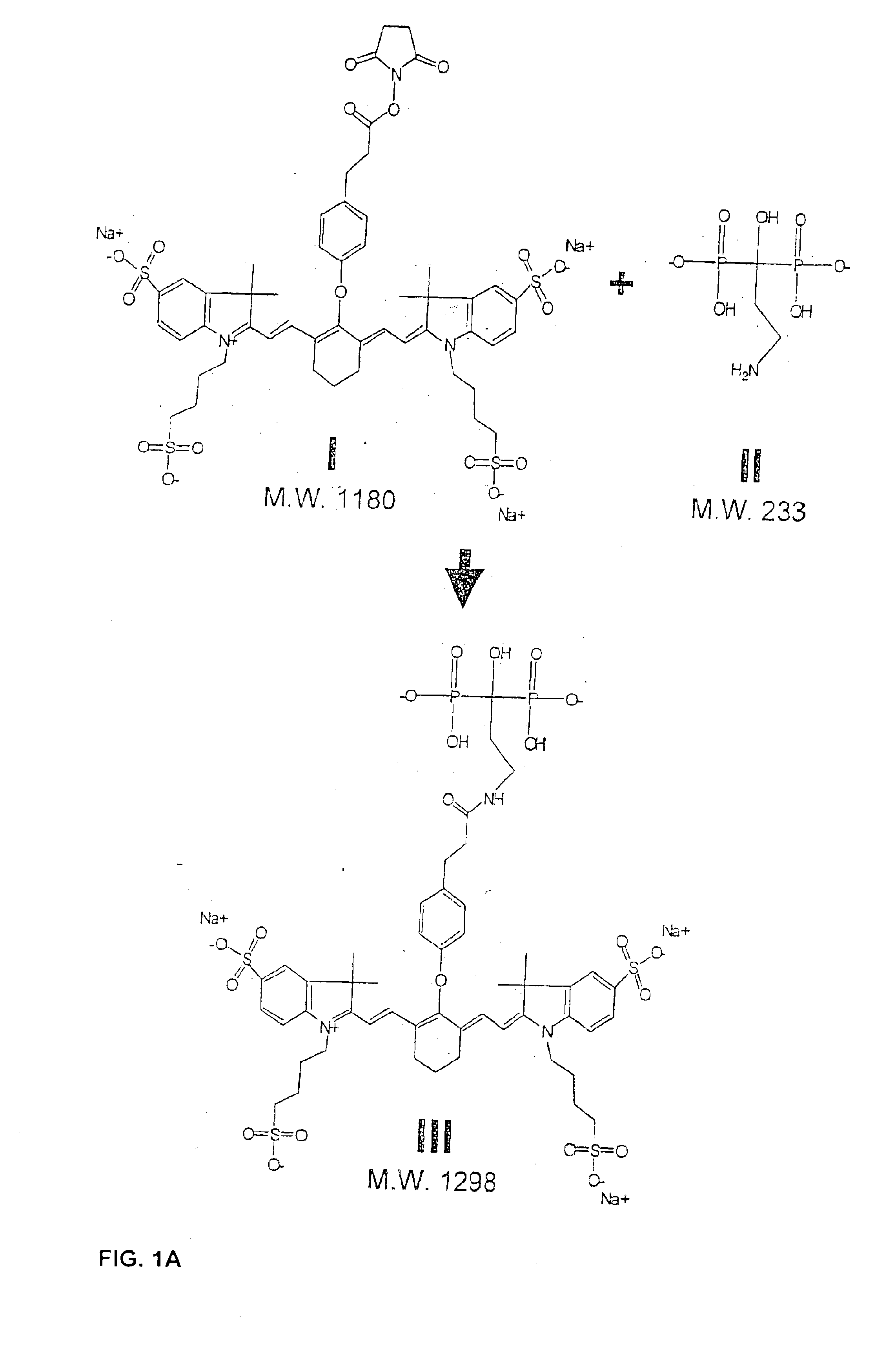
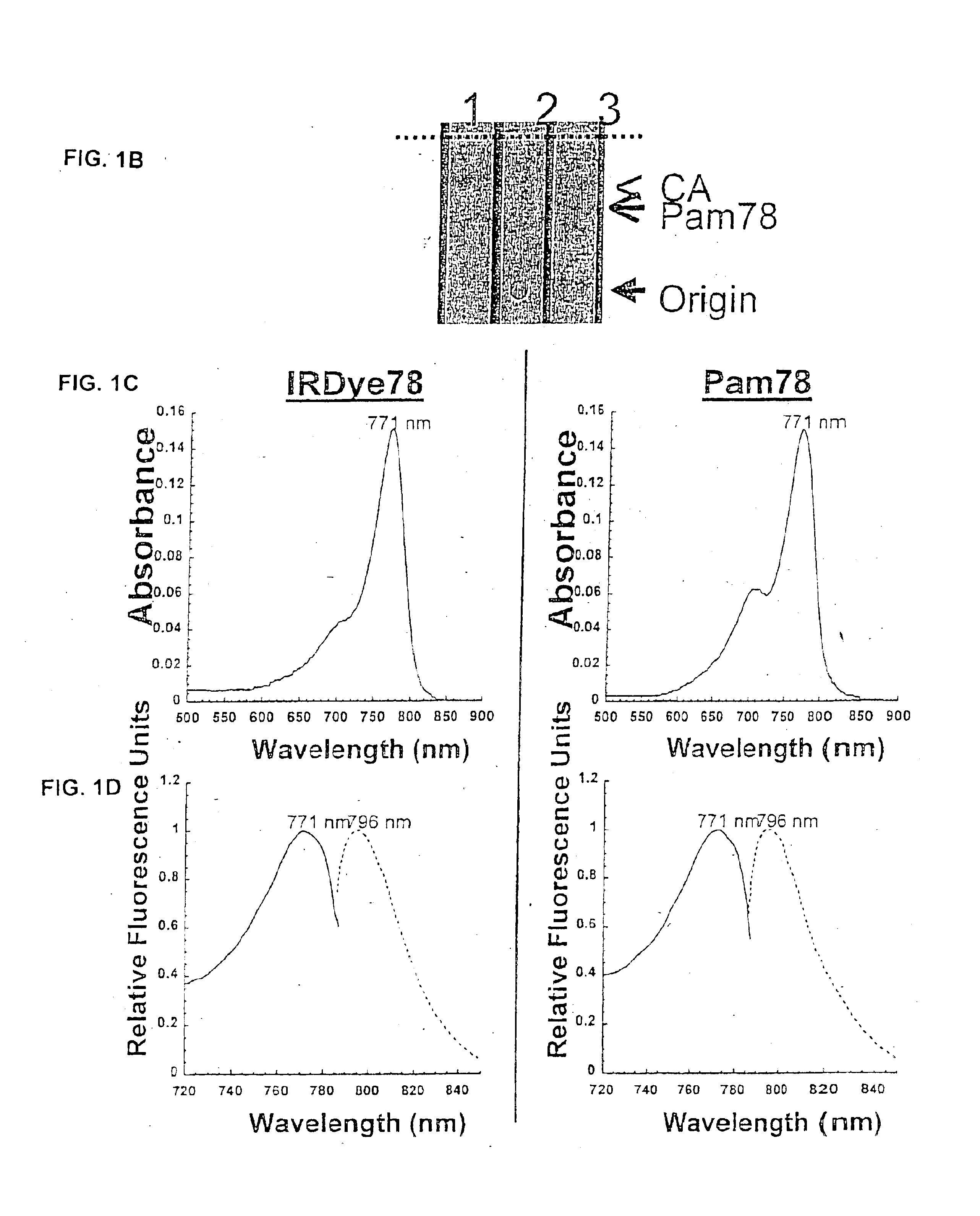
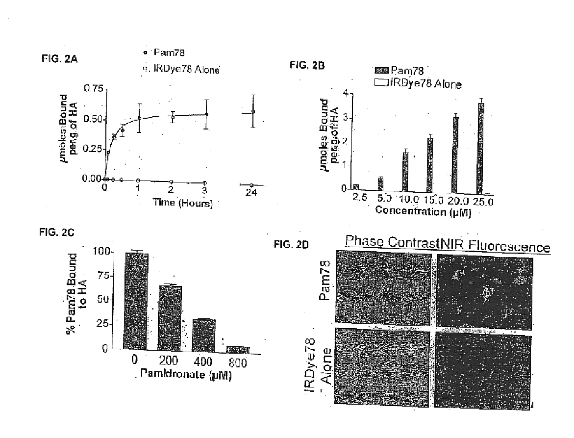
The present invention provides methods and compositions for improved imaging of tissues which include hydroxyapatite. In particular, the present invention provides fluorescent bisphosphonate derivatives that exhibits rapid and specific binding to hydroxylapatite in vitro and in vivo.

Certain preferred embodiments are directed to the use of bisphosphonates coupled to near-infrared fluorescent dyes to generate contrast agents that retain high affinity and rapid binding to hydroxyapatite. Relative to visible light flu...


PUM

PropertyMeasurementUnit
quantum efficiencyaaaaaaaaaa
excitation and emission spectraaaaaaaaaaa
fluorescentaaaaaaaaaa

Abstract

The present invention is directed to a non-isotopic methods for the in vitro and in vivo detection of hydroxyapatite-positive cells and structures.

Description

BACKGROUND OF THE INVENTIONThe development and maintenance of the vertebral skeleton is a complex process, but in simplest terms represents a balance between osteoblast-induced mineralization and osteoclast-induced demineralization. Osteoblast-like cells are also present in the vascular wall and participate in the earliest manifestations of atherosclerosis. Hydroxyapatite (HA; Ca10(PO4)6(OH)2, also know as hydroxylapatite) is the major mineral product of osteoblasts and calcifying vascular cells and binds naturally occurring pyrophosphates and phosphonates with high affinity. Osteoblastic activity occurs at sites of new bone formation, i.e., sites of deposition of hydroxyapatite. New bone formation occurs after fracture, at sites of bony infections, and especially at the sites of certain cancer metastases (e.g., prostate cancer metastases).At the present time, osteoblastic activity is detected in vivo using radionuclides and SPECT imaging. For instance, a common technique is the “bo...

Claims


Application Information

Patent Timeline
Patent Type & Authority Patents(United States)
IPC IPC(8): A61K49/00C07F9/572
CPCA61K49/0032C07F9/5728A61K49/005A61P43/00
Inventor FRANGIONI, JOHN V.
Owner BETH ISRAEL DEACONESS MEDICAL CENT INC
Why Eureka
Try Eureka