
X-ray computed tomography apparatus and picture quality simulation apparatus

a computed tomography and simulation apparatus technology, applied in tomography, applications, instruments, etc., can solve the problems of difficult to grasp the value of image sd, difficult operation, and inability to adjust the scan conditions suitably

Active Publication Date: 2006-04-18
TOSHIBA MEDICAL SYST CORP
View PDF7 Cites 40 Cited by
  • Summary
  • Abstract
  • Description
  • Claims
  • Application Information

AI Technical Summary

Benefits of technology

[0008]It is an object of the present invention to provide an X-ray computed tomography apparatus and a picture quality simulation apparatus which can set scan conditions more suitably.

Problems solved by technology

A reconstructed image inevitably contains image noise.
As disclosed in Jpn. Pat. Appln. KOKAI Publication No. 11-235334, it is difficult to grasp the value of image SD only by inputting many condition items such as an imaging slice thickness, tube voltage, and tube current.
This is not an easy operation.
It is therefore difficult to suitably set scan conditions.

Method used


Image

  • X-ray computed tomography apparatus and picture quality simulation apparatus
  • X-ray computed tomography apparatus and picture quality simulation apparatus
  • X-ray computed tomography apparatus and picture quality simulation apparatus

Examples


Embodiment Construction

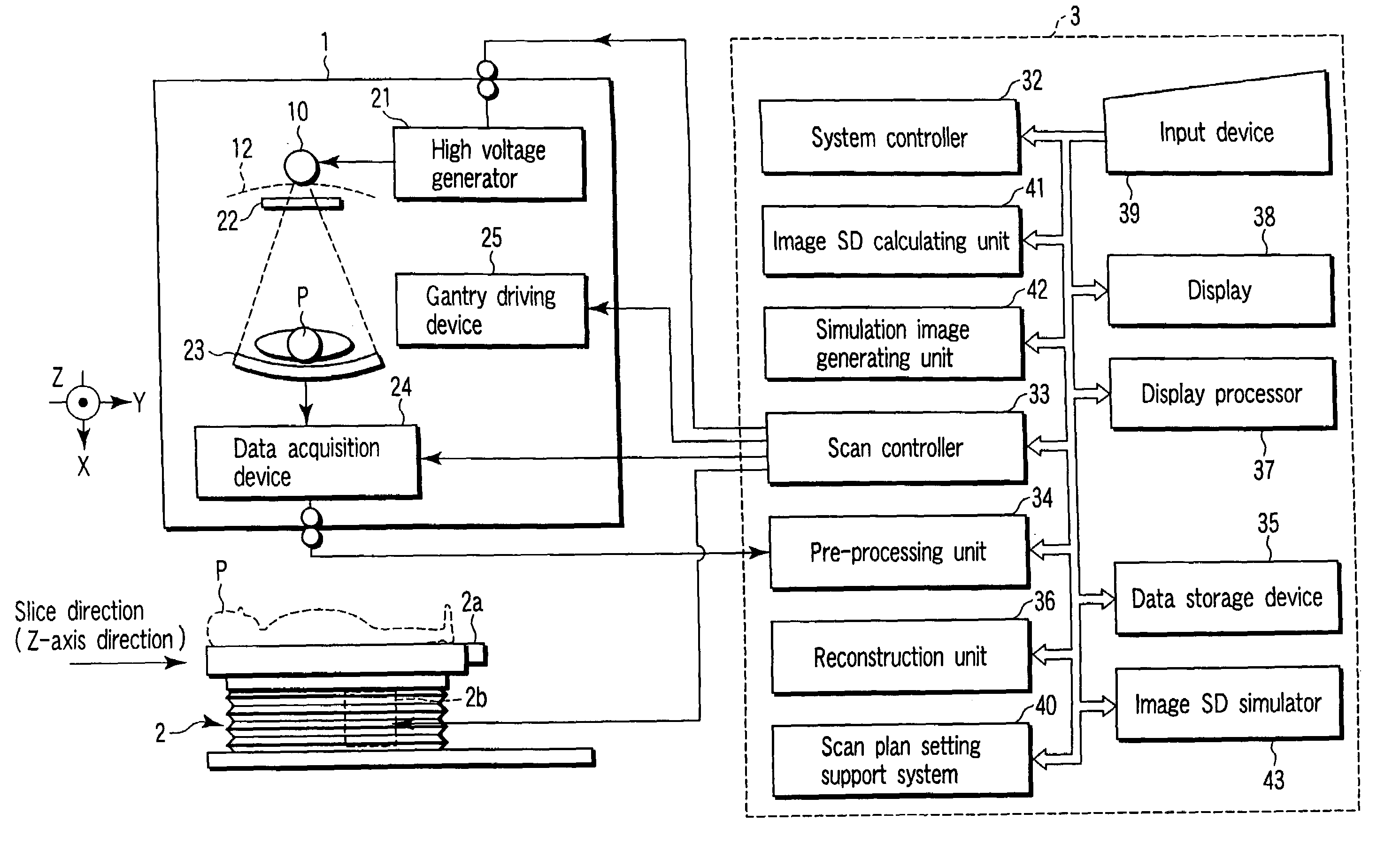
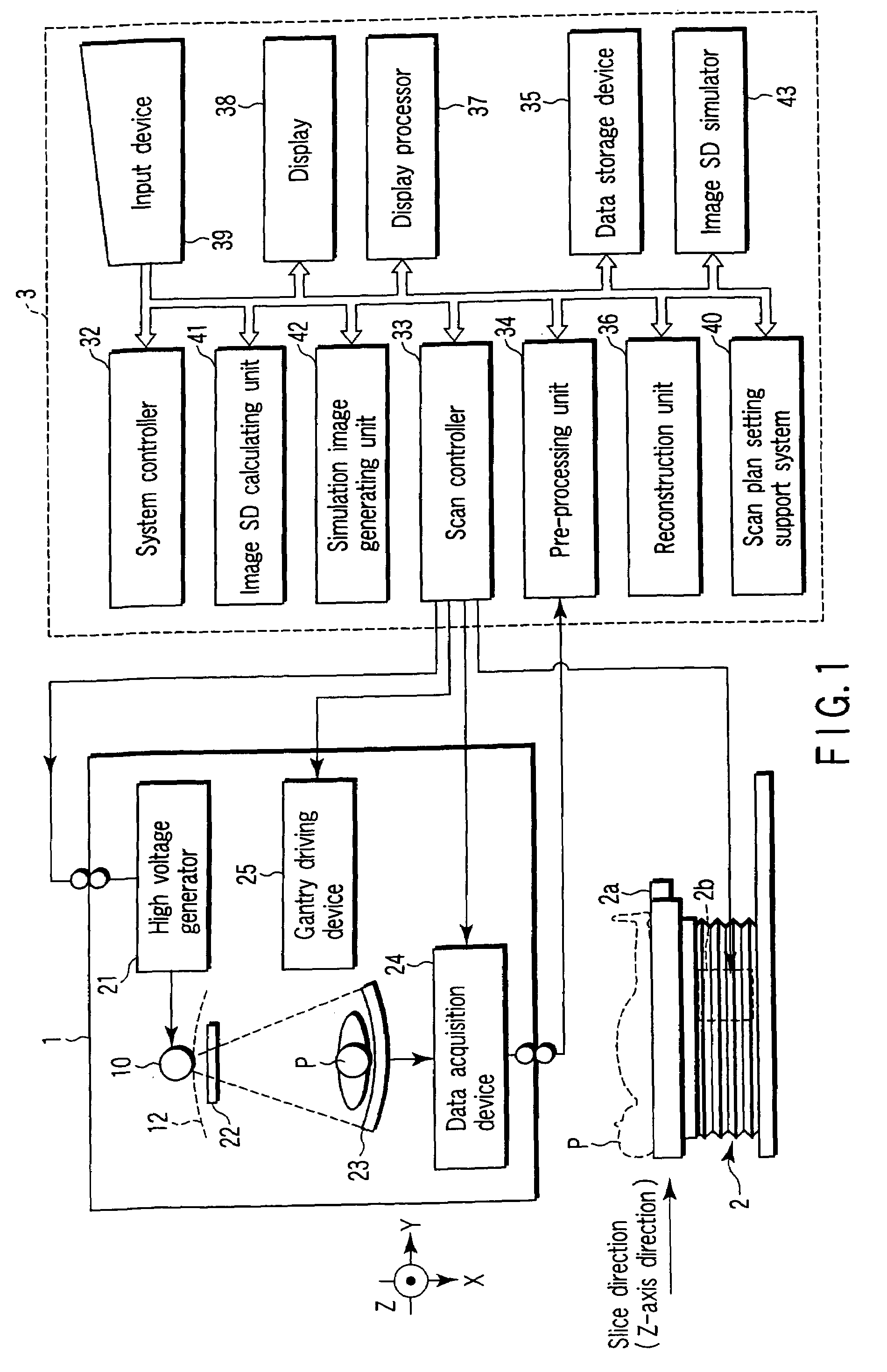
[0021]An X-ray computed tomography apparatus according to an embodiment of the present invention will be described below with reference to the views of the accompanying drawing. Note that X-ray computed tomography apparatuses include various types of apparatuses, e.g., a rotate / rotate-type apparatus in which an X-ray tube and X-ray detector rotate together around a subject to be examined, and a stationary / rotate-type apparatus in which many detection elements arrayed in the form of a ring, and only an X-ray tube rotates around a subject to be examined. The present invention can be applied to either type. In this case, the rotate / rotate type, which is currently the mainstream, will be exemplified. In order to reconstruct one-slice tomographic image data, projection data corresponding to one rotation around a subject to be examined, i.e., about 360°, is required, or (180°+ view angle) projection data is required in the half scan method. The present invention can be applied to either o...


PUM


Abstract

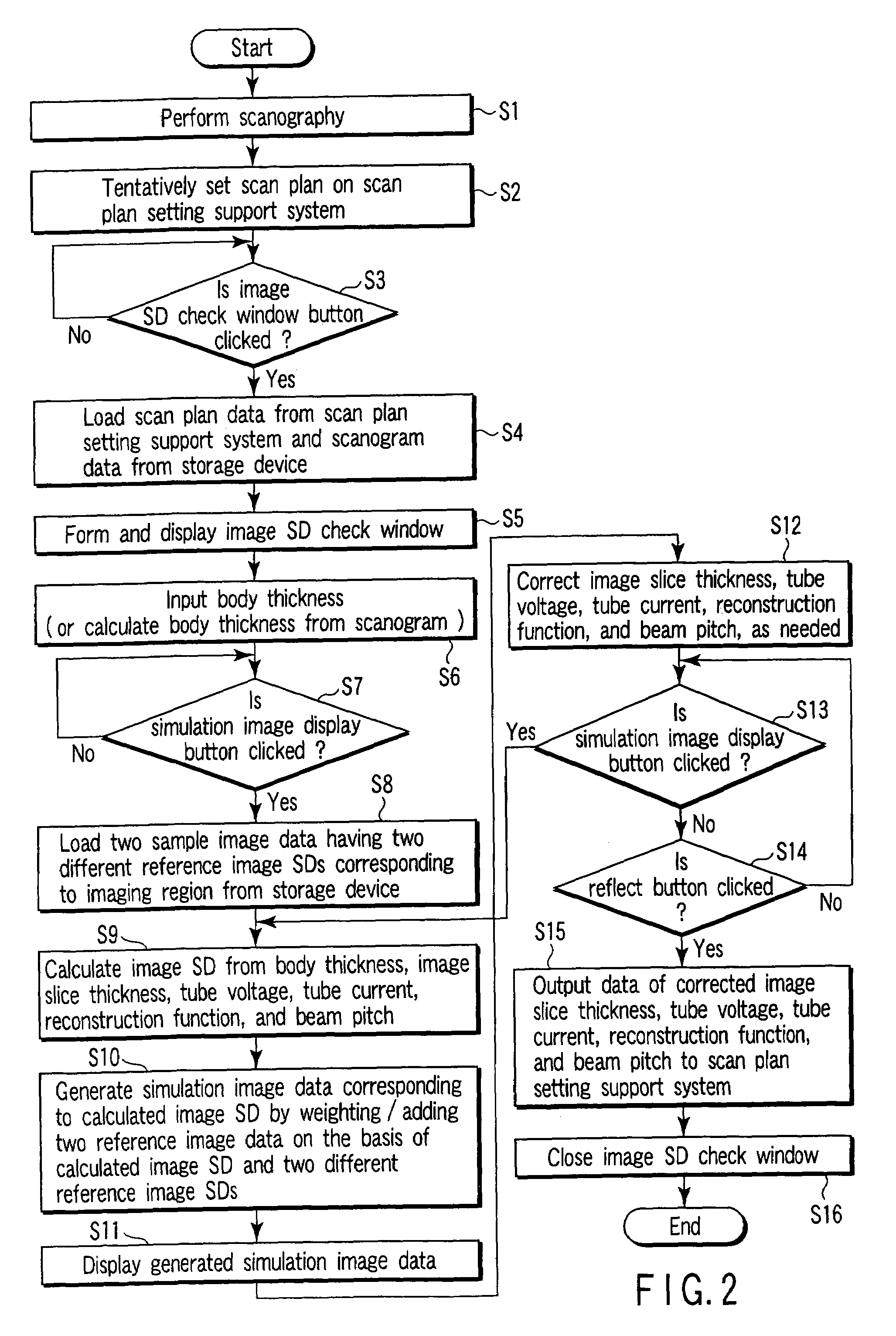
An X-ray computed tomography apparatus according to this invention includes a gantry (1) which scans an imaging target region of a subject to be examined in accordance with scan conditions, a reconstruction unit (36) which reconstructs image data from projection data, a scan plan setting support system (40) which sets scan conditions, an image SD calculating unit (41) which calculates an image SD associated with an index of picture quality on the basis of the set scan conditions, a data storage device (35) which stores sample image data having a reference value of an image SD, a simulation image generating unit (42) which generates simulation image data corresponding to the calculated image SD from the sample image data on the basis of the calculated image SD and the reference value of the image SD, and a display (38) which displays the generated simulation image data.

Description

CROSS-REFERENCE TO RELATED APPLICATIONS[0001]This application is based upon and claims the benefit of priority from the prior Japanese Patent Application No. 2003-131613, filed May 9, 2003, the entire contents of which are incorporated herein by reference.BACKGROUND OF THE INVENTION[0002]1. Field of the Invention[0003]The present invention relates to an X-ray computed tomography apparatus and picture quality simulation apparatus.[0004]2. Description of the Related Art[0005]As is known, an X-ray computed tomography apparatus is designed to obtain an image (tomographic image) by calculating (reconstructing) the X-ray absorption coefficient of a tissue such as an organ on the basis of the amount of X-rays absorbed in a subject to be examined as an index called a CT value with reference to the X-ray absorption coefficient of water. A reconstructed image inevitably contains image noise. Image noise is typically defined with reference to a variation in the CT value of a homogeneous phanto...

Claims


Application Information

Patent Timeline
Patent Type & Authority Patents(United States)
IPC IPC(8): A61B6/00A61B6/03
CPCA61B6/032A61B6/463A61B6/544A61B6/583A61B6/542Y10S378/901
Inventor TSUKAGOSHI, SHINSUKE
Owner TOSHIBA MEDICAL SYST CORP
Learn More