System for computerized processing of chest radiographic images
What is AI technical title?
AI technical title is built by Patsnap AI team. It summarizes the technical point description of the patent document.
a computerized processing and chest radiographic technology, applied in the field of chest radiographic image computerized processing system, can solve the problems of difficult chest radiograph detection task for radiologists, inability to apply temporal subtraction technique in absence of previous chest radiograph, etc., to achieve enhanced asymmetrical opacities, improved subtraction images, and improved sensitivity
Inactive Publication Date: 2006-05-09
ARCH DEVMENT
View PDF13 Cites 39 Cited by
Summary
Abstract
Description
Claims
Application Information
AI Technical Summary
This helps you quickly interpret patents by identifying the three key elements:
Problems solved by technology
Method used
Benefits of technology
Benefits of technology
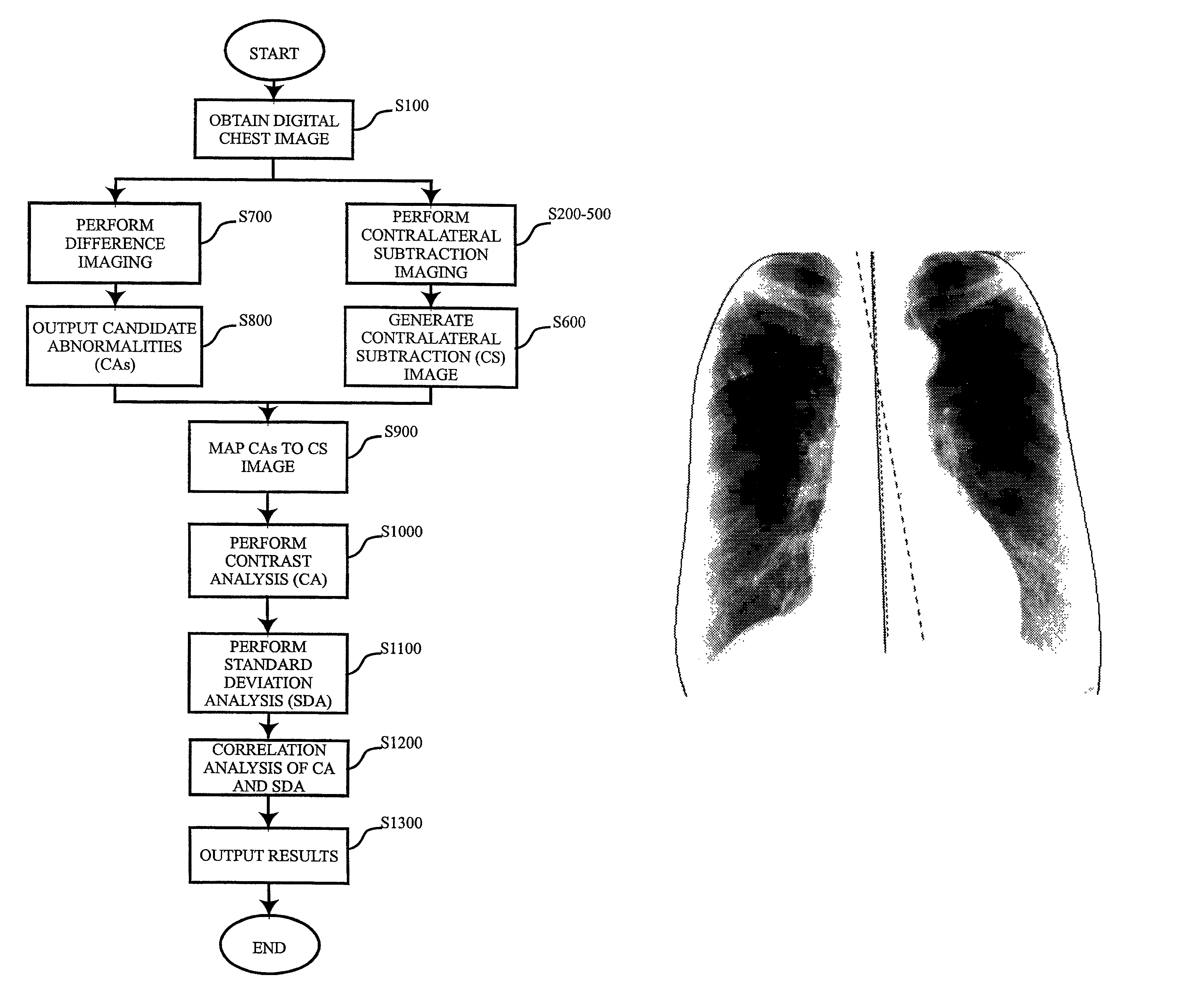
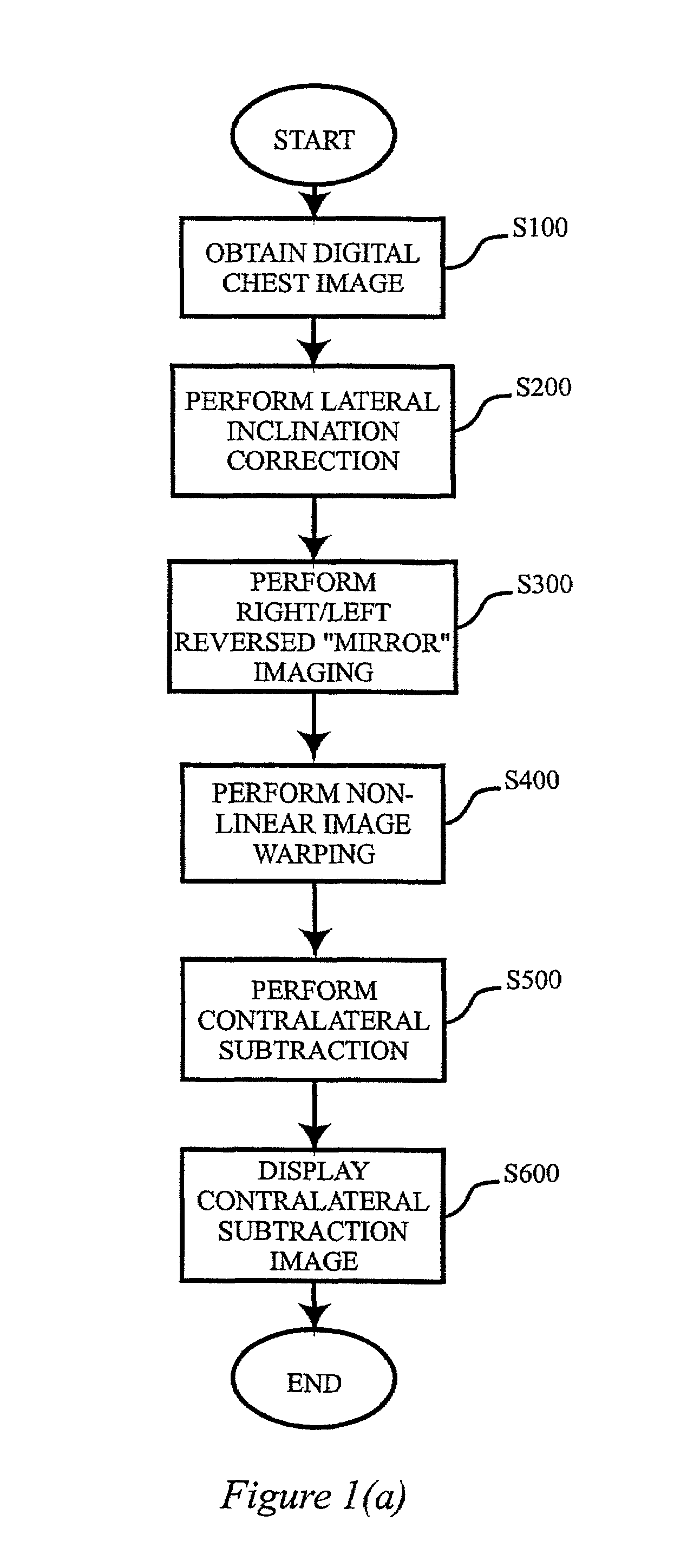
[0017]Thus, according to the present invention, a new contralateral subtraction technique based on one posteroanterior (PA) chest image has been developed. Since the rib structure is nearly symmetrical, the chest image of the right peripheral lung is generally similar to that of the left lung. Taking advantage of this, the technique includes lateral inclination correction by rotating and shifting the original chest image so that the midline of the thorax is aligned with the vertical centerline of the original chest image, lateral reversal of the rotated image to produce a reversed “mirror” image, warping of the mirror image, and subtraction of the warped mirror image from the original image to derive the contralateral subtraction image. Thereafter, additional processing techniques can be successively applied to the initial contralateral subtraction image to acquire improved subtraction images. In the technique, a contralateral subtraction image can be obtained by subtracting the right / left reversed “mirror” image from a single chest image. Similar to the temporal subtraction technique, the contralateral subtraction scheme can cancel out most of symmetrical skeletal structures and enhance asymmetrical opacities, thus demonstrating subtle abnormalities more clearly. On the other hand, unlike the temporal subtraction technique, a subtraction image can be obtained whenever a single PA chest image is available. Therefore, the contralateral subtraction technique can be of significant clinical importance in some cases.
Problems solved by technology
Detection of early lung cancers on chest radiographs is a difficult task for radiologists, because subtle lesions tend to be low in contrast and can overlap with ribs and clavicles.
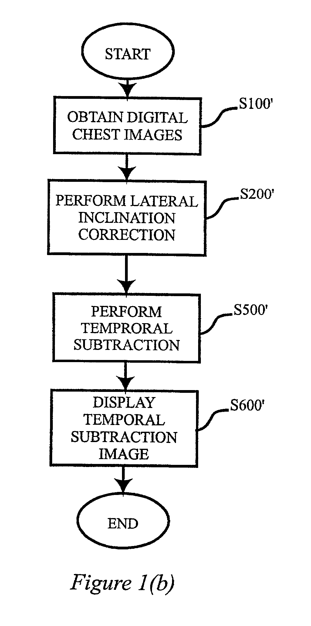
[2] However, the temporal subtraction technique is not applicable in the absence of a previous chest radiograph.
Method used
Image
Click on the blue label to locate the original text in one second.
Reading with bidirectional positioning of images and text.
Examples
Embodiment Construction
[0034]The chest images used in development of the present invention consist of 50 normals and 50 abnormals with solitary lung nodules which were selected from 247 chest images in the Japanese Standard Digital Image Database developed by the Japanese Society of Radiological Technology. [3] The images were digitized with a 0.175 mm pixel size, a matrix size of 2048×2048 and 12 bits gray levels. However, the matrix size was reduced to 512×512 by subsampling of the original image data, and the number of gray levels was decreased to 10 bits.
[0035]Referring now to the drawings, wherein like reference numerals designate identical or corresponding parts throughout the several views, and more particularly to FIG. 1(a) thereof, there is illustrated a top-level block diagram of a contralateral subtraction technique according to the present invention.
[0036]First of all, for an obtained PA chest image in step S100, the lateral inclination which may be caused by an improper patient positioning is...
PUM
Abstract
A method, system and computer readable medium for computerized processing of chest images including obtaining a digital first image of a chest (S100); producing a second image which is a mirror image (S300) of the first image; performing image warping on one of the first and second images to produce a warped image (S400) which is registered to the other of the first and second images; and subtracting the warped image from the other image to generate a subtraction image (S600). Another embodiment includes obtaining a digital first image of the chest of a subject; detecting ribcage edges on both sides of the lungs in the first chest image; determining average horizontal locations of the left and right ribcage edges at plural vertical locations; fitting the determined average horizontal locations to a straight line to derive a midline; rotating the chest image so that the midline is vertical; and shifting the rotated image to produce a lateral inclination corrected (S200) second image with the midline centered in the lateral inclination corrected image.
Description
STATEMENT REGARDING FEDERALLY SPONSORED RESEARCH[0001]The present invention was made in part with U.S. Government support under USPHS grant numbers CA62625 and CA64370 (National Institute of Health). The U.S. Government has certain rights in the invention.BACKGROUND OF THE INVENTION[0002]1. Field of the Invention[0003]The present invention relates generally to a computerized method and system provided to aid radiologists in detection of abnormalities, such as lung nodule, pneumothorax, pneumonia, and bulla, in chest radiographs.[0004]The present invention also generally relates to computerized techniques for automated analysis of digital images, for example, as disclosed in one or more of U.S. Pat. Nos. 4,839,807; 4,841,555; 4,851,984; 4,875,165; 4,907,156; 4,918,534; 5,072,384; 5,133,020; 5,150,292; 5,224,177; 5,289,374; 5,319,549; 5,343,390; 5,359,513; 5,452,367; 5,463,548; 5,491,627; 5,537,485; 5,598,481; 5,622,171; 5,638,458; 5,657,362; 5,666,434; 5,673,332; 5,668,888; 5,740,268...
Claims
Application Information
Patent Timeline
Application Date:The date an application was filed.
Publication Date:The date a patent or application was officially published.
First Publication Date:The earliest publication date of a patent with the same application number.
Issue Date:Publication date of the patent grant document.
PCT Entry Date:The Entry date of PCT National Phase.
Estimated Expiry Date:The statutory expiry date of a patent right according to the Patent Law, and it is the longest term of protection that the patent right can achieve without the termination of the patent right due to other reasons(Term extension factor has been taken into account ).
Invalid Date:Actual expiry date is based on effective date or publication date of legal transaction data of invalid patent.

