Systems and graphical user interface for analyzing body images

a graphical user interface and system technology, applied in healthcare informatics, local control/monitoring, instruments, etc., can solve the problems of inability to reliably detect small nodules, increased probability that the radiologist will miss a potential nodule in their analysis of the image dataset, and appearance may be obscured

Inactive Publication Date: 2006-10-31
MERATIVE US LP
View PDF58 Cites 133 Cited by
  • Summary
  • Abstract
  • Description
  • Claims
  • Application Information

AI Technical Summary

Benefits of technology

[0009]The present invention provides graphical user interfaces and systems that allow an operator to localize and analyze lung nodules in a baseline and follow up lung CT or MRI scan.
[0012]The nodule information can be stored and later transferred to a follow up image scan taken at t2 to ease the localization of the previously localized nodules in the follow-up scans. The stored nodule information provides a method that allows the operator to quickly determine if the nodules localized in the baseline scan have increased in size, the amount of increase, and the like. Additionally, the present invention can also allow the operator to quickly locate nodules—either manually or automatically with an analysis algorithm—that were not located in the baseline scan.
[0013]Typically, the graphical user interfaces of the present invention can be used to analyze thoracic images to track potential lung nodules over a time period. An operator can use the composite image to track the potential nodules (if any) in the first body image and the second body image. The composite image can provide the user a visual indication of any change in surface area, pixel area, or volume of the potential lung nodule.

Problems solved by technology

Unfortunately, such large amounts of data for each patient increases the probability that the radiologist will miss a potential nodule in their analysis of the image dataset, i.e., a “false negative.” Tumors may be too small to be reliably detected, or their appearance may be obscured by the surrounding tissues such as vessels.
In their early stages of development, malignant lung tumors may not be detected even upon careful inspection of the image dataset.
It is generally believed that early detection of cancer is beneficial, but in the case of lung cancer this is not established because of the recentness of the technique.
Conventional imaging systems and methods do not provide an efficient way to determine if the nodules have increased in size, stayed the same, or the like.

Method used


Image

  • Systems and graphical user interface for analyzing body images
  • Systems and graphical user interface for analyzing body images
  • Systems and graphical user interface for analyzing body images

Examples


Embodiment Construction

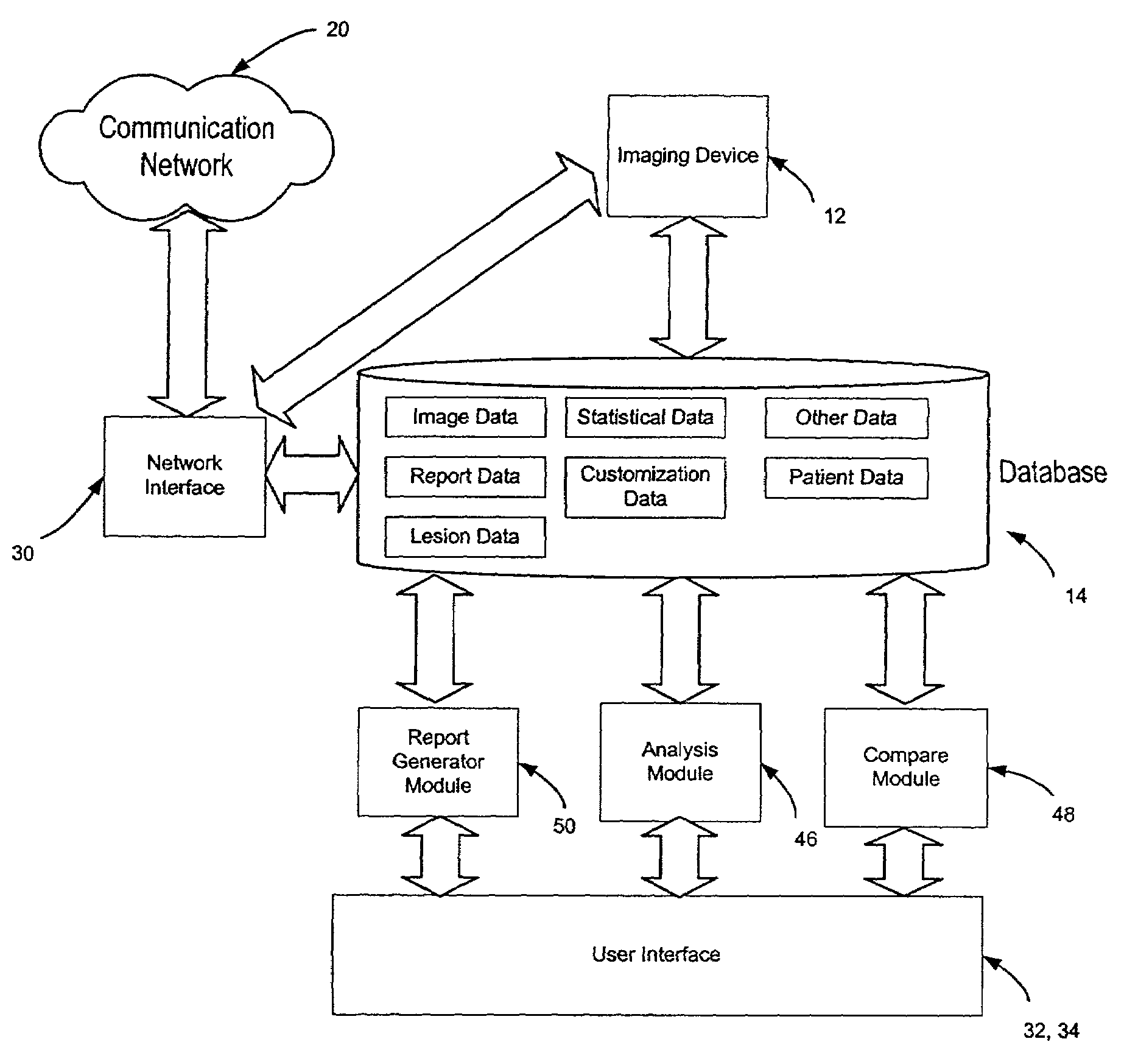
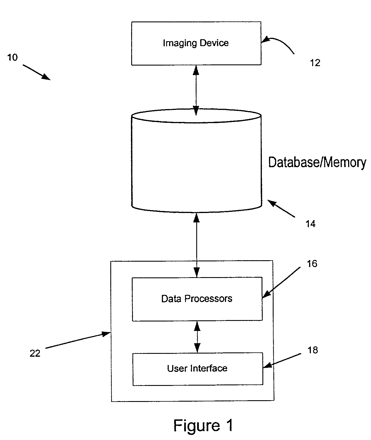
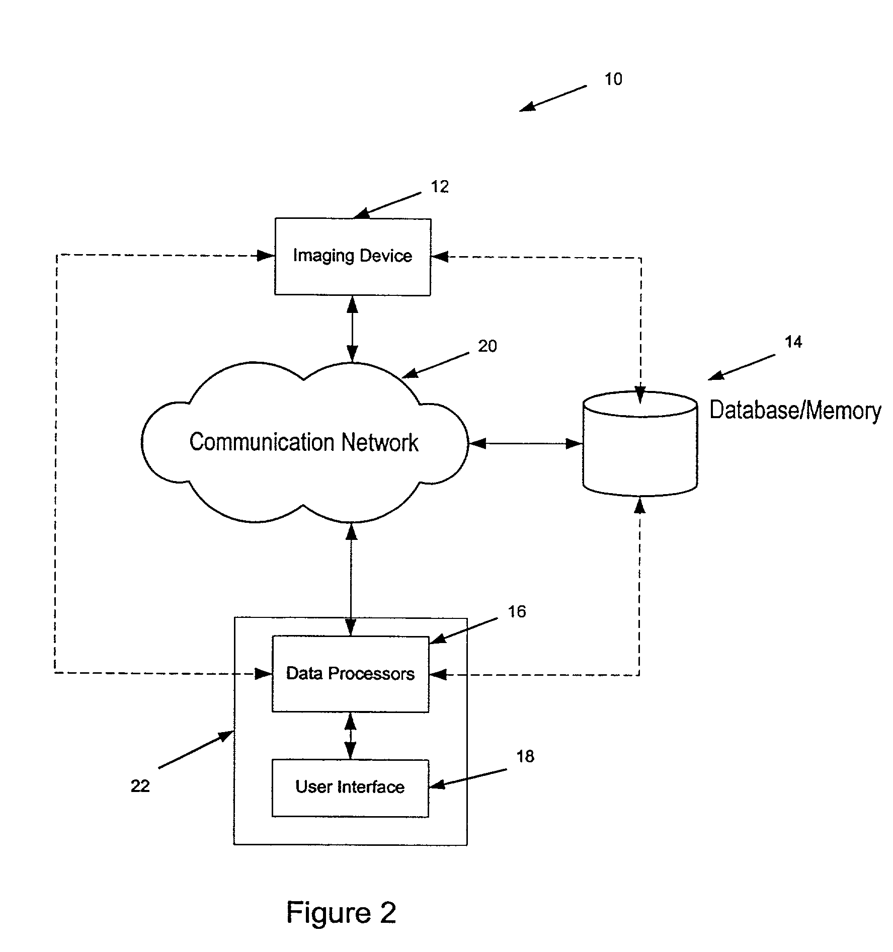
[0042]The present invention provides systems, software code, graphical user interfaces, and methods for displaying and analyzing lung CT or MRI image datasets of a patient. The lung datasets can be analyzed to map, track, and analyze the nodules in a series of lung slice images or image scans, as well as record other lung and chest abnormalities.

[0043]A lung slice image can be displayed on a user interface display for analysis by a radiologist or other operator. The methods of the present invention allows the radiologist to locate and map out the tumors, nodules, or lesions (hereinafter referred to as “nodules”) that are both manually localized and / or automatically localized by the software of the present invention. The mapped nodules can be segmented and have its volume and other dimensions ascertained. Such nodule information can then be transferred onto a lung report, if desired.

[0044]Exemplary embodiments of the present invention may allow an operator to compare a first, baselin...


PUM


Abstract

Systems and graphical user interfaces for analyzing body images. In an exemplary embodiment, the present invention provides a graphical user interface having a display coupled to a microprocessing device and a memory device. The graphical user interface has an electronic representation of a first body image and a second body image and an electronic map representing the position of nodules on the first body image and second body image.

Description

CROSS-REFERENCES TO RELATED APPLICATIONS[0001]The present application is related to U.S. patent application Ser. No. 09 / 908,432, filed herewith and entitled “Customizable Lung Report Generator” and U.S. patent application Ser. No. 09 / 908,466, filed concurrently herewith and entitled “Methods and Systems for Generating a Lung Report,” each of which is incorporated by reference.BACKGROUND OF THE INVENTION[0002]The present invention relates generally to systems, methods, software, and graphical user interfaces for displaying and analyzing body images and for generating organ reports. More particularly, the present invention relates to graphical user interfaces and systems for analyzing one or more thoracic CT datasets to track and analyze lung nodules and other lung parameters.[0003]Lung cancer is one of the most common forms of cancer among both men and women. Advances in medical imaging, such as CT and MRI scanning, have made it possible to localize and track early stage nodules that...

Claims


Application Information

Patent Timeline
Patent Type & Authority Patents(United States)
IPC IPC(8): G06K9/00G06F19/00
CPCG06F19/321G06F19/3406G06F19/3487G16H40/63G16H15/00G16H30/20
Inventor KAUFMAN, LEONMINEYEV, MIKHAILPOWERS, SHELLEYGOLDHABER, DAVID
Owner MERATIVE US LP
Why Eureka
Try Eureka