Object identifying system for segmenting unreconstructed data in image tomography

an object identification system and image tomography technology, applied in image enhancement, image analysis, instruments, etc., can solve the problems of contaminating later data processing, affecting and affecting the quality of image tomography, so as to maintain the statistical independence of object data

Inactive Publication Date: 2007-02-13
TIES
View PDF8 Cites 4 Cited by
  • Summary
  • Abstract
  • Description
  • Claims
  • Application Information

AI Technical Summary

Benefits of technology

[0015]The invention, in one or more of its preferred embodiments, simplifies and expedites the selection and isolation of objects within the unreconstructed tomographic projection data for such purposes as maintaining the statistical independence of the object data, removing obscuring objects from image reconstructions, or providing composite image reconstructions based on different segmentations of the projection data. Instead of identifying objects by selecting isolated portions of their traces throughout a series of sinograms corresponding to the transaxial slices in which the object appears, objects can be identified by selecting isolated full views of the objects within the set of data frame images captured by the gamma cameras or other detectors at different angular positions around the target body. Object approximations can be made for the remaining data frame images to complete a three-dimensional definition of the object, which can be referenced either to the transaxial slices of the target body in which the object appears in cross section or to the series of sinograms in which the object appears as a sinusoidal trace through an angularly-sensitive arrangement of the projection data.
[0016]Once the object is spatially identified, an appropriate portion of the radiation counts from the projection data can be assigned to the object. Removing the selected object's contribution to the radiation counts enables an image of the remaining projection data to be reconstructed without the influence of the selected object on the reconstructed image. The deleterious influence of high-intensity objects for producing unwanted artifacts and blurring can be mitigated by reconstructing images based on projection data that has been segmented to remove the high-intensity object's contribution to the radiation counts. Conversely, the projection data pertaining to the selected object can be retained, and the remaining radiation counts attributable to other objects or features can be discounted for reconstructing an image of the selected object on its own scale of intensities.
[0018]The segmentation process involves two steps: (a) the creation of segmentation masks for spatially isolating selected objects and (b) the attribution of appropriate radiation counts to the selected objects within their defined space. A number of different mask-creating techniques can be applied to the camera data frames to achieve the desired spatial definitions of selected objects; and other techniques, based on aspects of geometric predictability in the sinogram presentation, can be used to appropriately assign radiation counts to the unique spaces occupied by the selected objects. The techniques enable the acquisition of quantitative time-sensitive measurements of the radiological activities of the selected objects, and this additional information can be presented in a graphic or numerical format for supplementing or further interpreting the reconstructed images.
[0021]Areas bounded by the outlines of the selected object within the data frames define a set of masks. The sizes of the mask areas can be adjusted in one or more directions, such as by changing the condition that is required to be satisfied for identifying a pixel as being within the outline of the object. A curve or other approximation can be fit through the interior points within the object outlines to identify other pixels within the outlines of the same object appearing in other of the data frames. The outlines of the object in the remaining data frames can be found by identifying pixels that are contiguous to the fitted pixels and that satisfy the condition for being within the outlines of the object. Alternatively, curves can be fit through the identified outlines of the object for defining boundaries of the circumferential traces within the set of sinograms. To accommodate overlapping objects and other data influences that obscure object outlines, limitations can be imposed upon the amount of change allowed between the outlines of angularly adjacent data frames. In addition, collecting collimated photons in 180° separated pairs of angular increments provides a redundancy supporting a choice among corresponding paired data frames in which the object outline is most easily distinguished from its surroundings.

Problems solved by technology

Thus, the voxel map produced in the reconstructed image cannot be considered as “raw data”, and further processing (e.g., for image enhancement or quantitative analysis) of the reconstructed data can encounter problems with unwanted filtering artifacts such as aliasing and the like.
Although resulting in a more accurate final image, such corrections also introduce filtering artifacts, which can further contaminate later data processing.
High intensity objects and sharp edges can also introduce a considerable amount of blur, including obscuring artifacts, into in the reconstructed image.
Frequency domain highpass filters are often used to remove the blur, but there is no perfect way to remove this blur and associated obscuring artifacts within the images.
If the intensities of some of the objects are much higher than others, then there will be more blur and hence more artifacts.
All of the above factors can severely limit the ability to accurately quantify the reconstructed data derived from SPET and PET studies.
In addition, the accuracy of quantitative data can be affected by other factors such as noise, sampling errors, detector characteristics, time variance of the radioactivity distribution during the period of acquisition, and the type of reconstruction algorithm and filtering used.
47–61; K A Blokland et al: “Quantitative analysis in single photon emission tomography.” However, none of the approaches are in widespread use and they fail to tackle a fundamental problem, namely, that of maintaining statistical independence of the sets of pixels or voxels of the reconstructed images (i.e., the projection data).
Distinguishing one object from another within the sinograms involves considerable manual and computer processing.
Thus, the segmentation of data is difficult, time consuming, and itself subject to variation depending on the accuracy with which the isolated sections of the object traces are identified and the rules for approximating of the remaining object data.

Method used


Image

  • Object identifying system for segmenting unreconstructed data in image tomography
  • Object identifying system for segmenting unreconstructed data in image tomography
  • Object identifying system for segmenting unreconstructed data in image tomography

Examples


Embodiment Construction

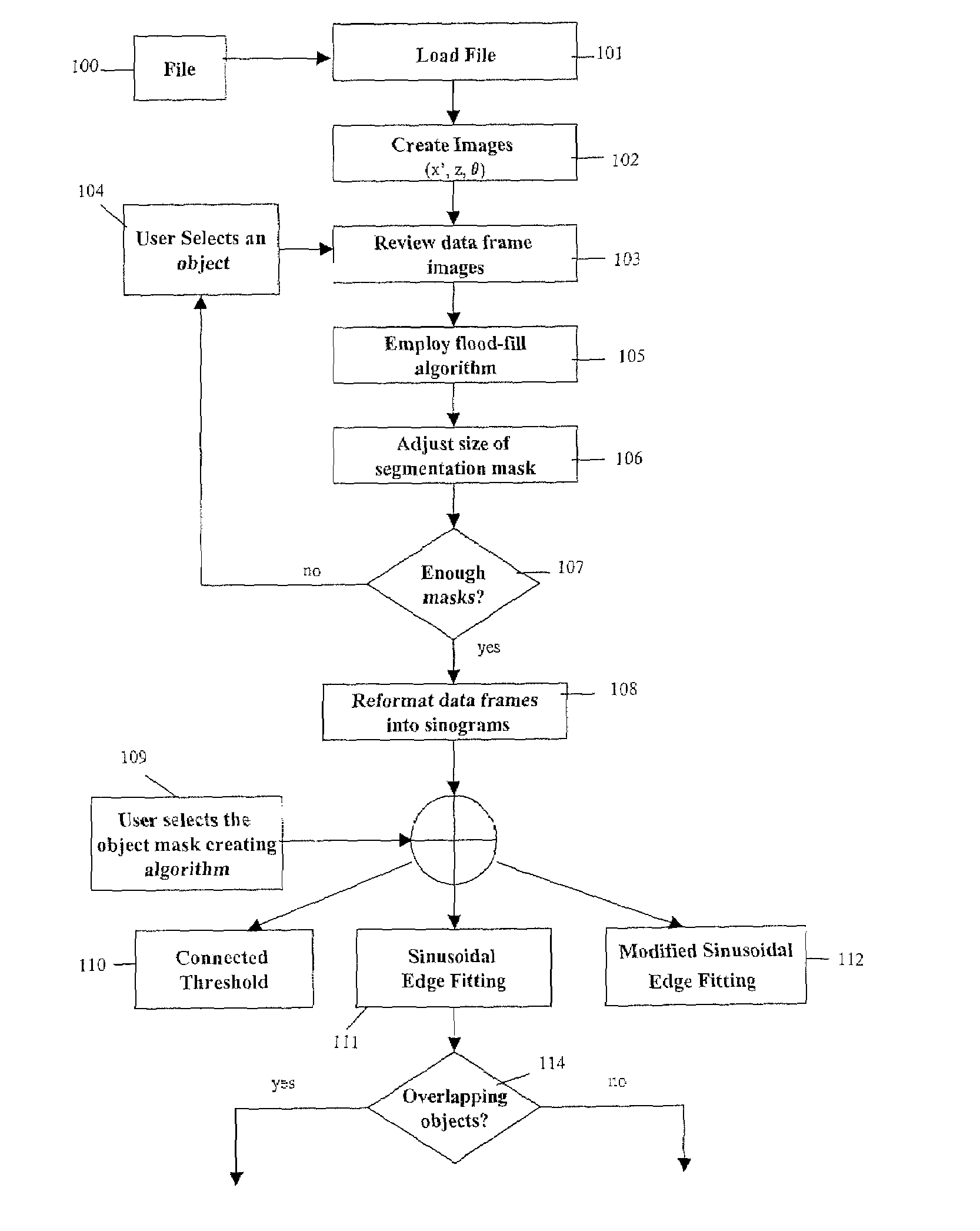
[0041]The invention, as described below, provides for isolating the contributions of radiologically distinguishable objects from unreconstructed tomographic projection data. For example, unwanted contributions of high intensity objects can be removed from the unreconstructed tomographic data so that cross-sectional images reconstructed from the data are not adversely affected by the artifacts and blur otherwise produced by the high intensity objects. Alternatively, the isolated contributions of radiologically distinguishable objects collected from the unreconstructed tomographic data can be separately quantified or otherwise analyzed to determine their physical and radiological characteristics independently of the setting from which they are extracted. Separate reconstructions can be performed based on the segmented data, including reconstructions of the isolated objects or their remaining settings. The reconstructions can produce images that are combinable, yet independently evalua...


PUM


Abstract

Tomographic projection data is segmented spatially by forming segmentation masks over the full outlines of objects appearing in isolation within selected data frame images. Further processing locates the object outlines in other data frames. Predictable effects of attenuation are used to attribute appropriate amounts of intensity to the segmented object space. Separate image reconstructions can be made of the segmented object data and the remaining projection data to independently optimize the reconstructions. Composite images can be made of the separate reconstructions.

Description

TECHNICAL FIELD[0001]The invention relates generally to the field of image tomography, and in particular to methods of identifying, locating or analyzing objects of interest as well as tracking and segmenting or removing selected objects in SPET (single-photon emission tomography) or PET (positron-emission tomography) studies on the raw tomographic data.BACKGROUND[0002]The methodologies of SPET and PET are described in detail in several standard textbooks on Nuclear Medicine. Broadly speaking, the methodologies involve constructing a three-dimensional map of radioactivity sources within an entity or target body and displaying relative levels of radioactivity emanating from each of a plurality of volume elements making up the entity. The radioactivity sources are generally gamma radiation emitters (radiotracers), which have been injected into the body of a patient or other target body, and these are detected by a gamma camera which images a plurality of views of the body as illustrat...

Claims


Application Information

Patent Timeline
Patent Type & Authority Patents(United States)
IPC IPC(8): G06T17/00
CPCG06T7/0083G06T11/008G06T2207/30004G06T2207/10108G06T2207/10104G06T7/12
Inventor RIAZ, UMAR
Owner TIES
Why Eureka
Try Eureka