Excisional biopsy devices and methods

a biopsy device and biopsy technique, applied in the field of excisional biopsy devices and methods, can solve the problems of excessively large amount of normal breast tissue removed, time-consuming multi-step process, and major threat to breast cancer, so as to minimize the size of the incision, minimize the complications of ecchymosis, hematoma formation, and breast edema, and efficiently and safely excise suspicious lesions.

Inactive Publication Date: 2007-12-04
ENCAPSULE MEDICAL
View PDF109 Cites 260 Cited by
  • Summary
  • Abstract
  • Description
  • Claims
  • Application Information

AI Technical Summary

Benefits of technology

[0021]It is, therefore, an object of the present invention to provide devices and methods to efficiently and safely excise suspicious lesions from the breast. It is also an object of the present invention to provide devices and methods that remove the entire lesion intact with the minimum amount of normal tissue surrounding the lesion needed to provide adequate margins. It is a further object of the present invention to provide devices and methods that provide hemostasis in the breast to minimize complications of ecchymosis, hematoma formation, and breast edema. It is another object of the present invention to provide methods and devices to provide intra-tissue ultrasonic guidance to provide real time, in situ monitoring of the procedure. A still further object is to provide devices and methods that allow the physician to minimize the size of the incision though which the procedure is performed and to leave an aesthetically acceptable scar on the breast.

Problems solved by technology

Breast cancer is a major threat and concern to women.
The conventional method for diagnosing, localizing and excising nonpalpable lesions detected by mammogramphy generally involves a time-consuming, multi-step process.
Obtaining good margins of normal tissue is extremely dependent upon the skill and experience of the surgeon, and often an excessively large amount of normal breast tissue is removed to ensure that the lesion is located within the specimen.
This increases the risk of post-operative complications, including bleeding and permanent breast deformity.
As 80% of breast biopsies today are benign, many women unnecessarily suffer from permanent scarring and deformity from such benign breast biopsies.
Fine needle aspiration is also very dependent upon the skill and experience of the operator and can result in a high non-diagnostic rate (up to about 83%), due to inadequate sample collection or preparation.
The stereotactic table requires awkward positioning of the patient and may be extremely uncomfortable.
The woman must lie prone during the entire procedure, which may be impossible for some patients.
This may be extremely difficult and uncomfortable for the patient if the lesion is very posterior near the chest wall or high towards the axilla.
Bleeding is generally not an issue with fine needle aspiration, but is a legitimate complication of the former two methods.
This causes increased post-procedural pain and delays healing.
However, documented instances of cancerous cells seeding locally into needle tracks exist.
The long-term consequences of neoplasm seeding into needle tracks are currently unknown, again because the techniques are relatively new.
The breast, however, may be torqued and distorted between the compression plates such that when the plates are removed after the biopsy, the large core track left behind may not be straight, but actually tortuous.
This can result in permanent breast deformity.
For the fine needle aspirate, core biopsy and vacuum-assisted devices, the incision is usually very small and the scar almost unappreciable.
Such a large incision often results in a non-aesthetically placed scar.
They are not, however, the final solution.
Also, the conventional biopsy devices do not consider margins in their excisions and if cancer is diagnosed, the patient must undergo a needle-localized breast lumpectomy to ensure that adequate margins are removed around the cancer.
Margins, moreover, cannot be assessed while the breast is being compressed.

Method used


Image

  • Excisional biopsy devices and methods
  • Excisional biopsy devices and methods
  • Excisional biopsy devices and methods

Examples


Embodiment Construction

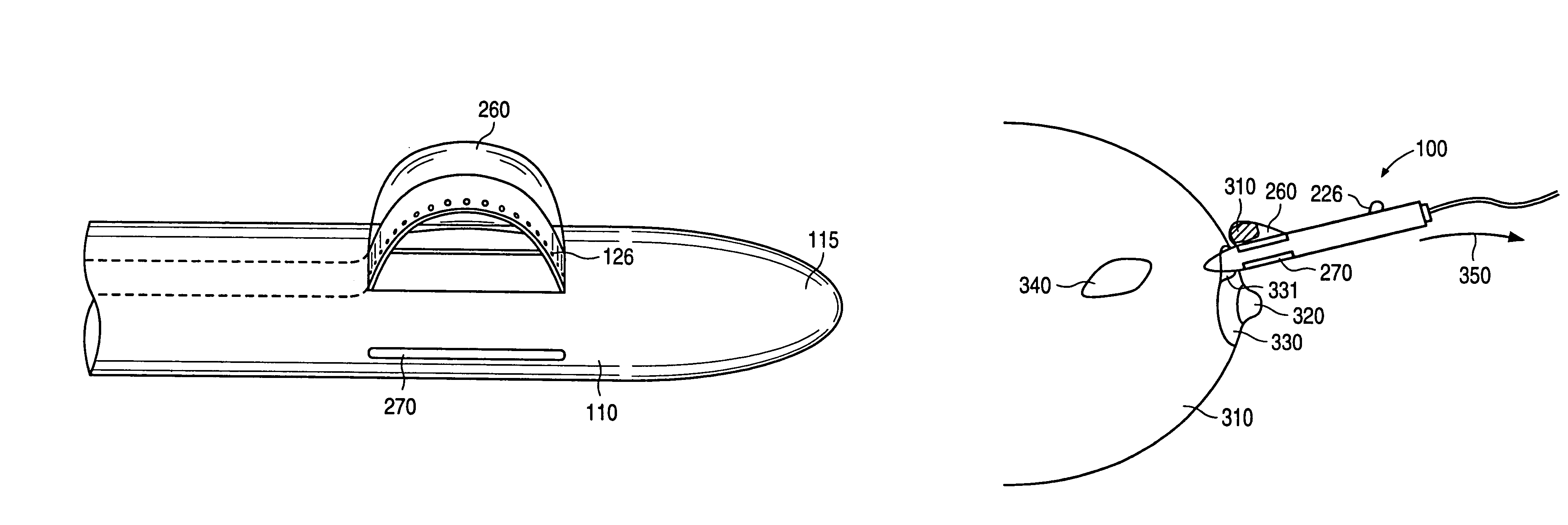
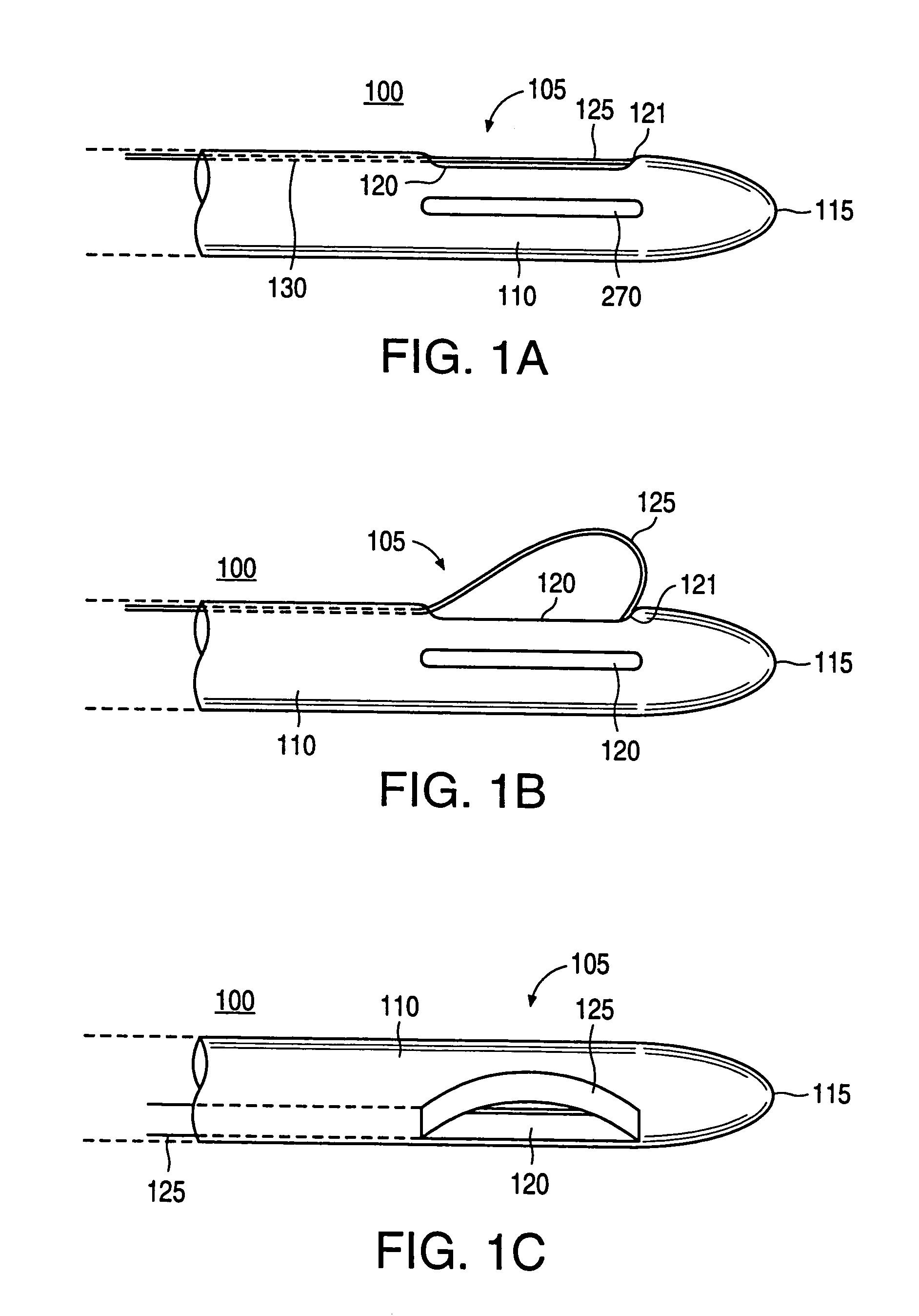
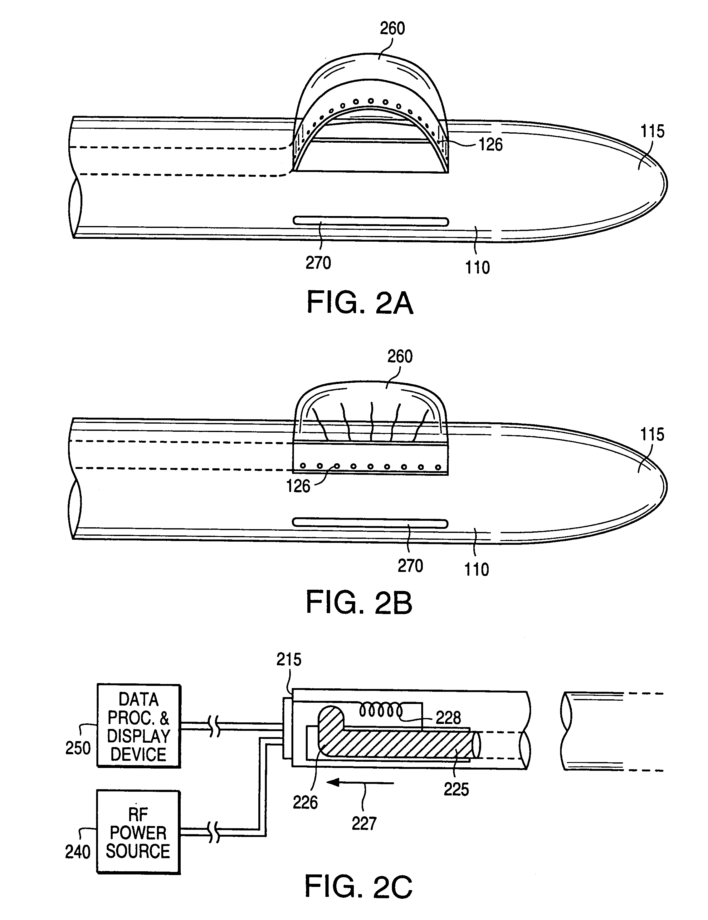
[0091]FIGS. 1A, 1B and 1C show an embodiment of the distal region 105 of the excisional biopsy device 100 according to the present invention. Considering FIGS. 1A, 1B and 1C collectively, the distal region 105 of the excisional biopsy device 100 includes a generally tubular member 110 having a generally tapered distal tip 115. The distal tip 115 is configured to penetrate soft tissue, such as breast tissue, lung tissue, liver tissue and the like. Preferably, therefore, the distal tip 115 and the distal region 105 of the excisional biopsy device 100 present a smooth, and relatively atraumatic profile to the soft tissue in which it is designed to penetrate. Alternatively, the tip 115 may be sharply pointed and / or may include an energy source (not shown) to facilitate cutting through the tissue. The tubular member 110 may be formed of rigid and hard plastic, or may be made of stainless steel, for example. Preferably, the tubular member 100 is used once and disposed of, for both safety ...


PUM


Abstract

An excisional biopsy device includes a tubular member having a window near a distal tip thereof; a cutting tool, a distal end of the cutting tool being attached near the distal tip of the tubular member, at least a distal portion of the cutting tool being configured to selectively bow out of the window and to retract within the window; and a tissue collection device externally attached at least to the tubular member, the tissue collection device collecting tissue excised by the cutting tool as the biopsy device is rotated and the cutting tool is bowed. An excision al biopsy method for soft tissue includes the steps of inserting a generally tubular member into the tissue, the tubular member including a cutting tool adapted to selectively bow away from the tubular member and an external tissue collection device near a distal tip of the tubular member; rotating the tubular member; selectively varying a degree of bowing of the cutting tool; collecting tissue severed by the cutting tool in the tissue collection device; and retracting the tubular member from the soft tissue. The tubular member may include an imaging transducer and the method may include the step of displaying information received from the transducer on a display device and the step of varying the degree of bowing of the cutting tool based upon the displayed information from the imaging transducer. Alternatively, the imaging transducer may be disposed within a removable transducer core adapted to fit within the tubular member.

Description

CROSS-REFERENCE TO RELATED APPLICATIONS[0001]The present application is a Continuation of commonly assigned and U.S. patent application Ser. No. 10 / 066,462 filed on Jan. 31, 2002, now U.S. Pat. No. 6,702,831 which is a Continuation of U.S. patent application Ser. No. 09 / 417,520, filed Oct. 13, 1999, now U.S. Pat. No. 6,423,081, which is a Divisional of U.S. Ser. No. 09 / 146,743, filed Sep. 3, 1998, now U.S. Pat. No. 6,022,362.BACKGROUND OF THE INVENTION[0002]1. Field of the Invention[0003]The present invention pertains to the field of soft tissue excisional biopsy devices and methods. In particular, the present invention relates to the field of devices and methods for excising suspicious lesions from soft tissue, such as breast tissue.[0004]2. Description of the Related Art[0005]Breast cancer is a major threat and concern to women. Early detection and treatment of suspicious or cancerous lesions in the breast has been shown to improve long term survival of the patient. The trend is, ...

Claims


Application Information

Patent Timeline
Patent Type & Authority Patents(United States)
IPC IPC(8): A61B5/00A61B1/00A61B10/02A61B8/12A61B10/00A61B10/04A61B10/06A61B17/22A61B17/28A61B17/32A61B17/3211A61B18/14G01B17/00G01B17/06
CPCA61B8/461A61B10/0275A61B10/04A61B17/320725A61B18/1482A61B8/085A61B10/0266A61B10/02A61B2017/00287A61B2018/00011A61B2018/1407A61B2018/1475A61B2019/5278A61B2019/528A61B2218/002A61B2090/3782A61B2090/3784
Inventor LEE, ROBERTAVETTER, JAMES W.
Owner ENCAPSULE MEDICAL
Why Eureka
Try Eureka