
Biopsy forceps device with transparent outer sheath

a biopsy forceps and transparent technology, applied in the field of endoscopic instruments, can solve the problems of compromising the subsequent performance and sterility of the biopsy forceps, human blood and tissue may become trapped in the biopsy forceps, and the re-sterilization process is not always effective at fully cleaning the biopsy forceps designed for single use, so as to prevent such reuse and confirm adequate cleaning

Active Publication Date: 2008-03-11
BOSTON SCI SCIMED INC
View PDF177 Cites 23 Cited by
  • Summary
  • Abstract
  • Description
  • Claims
  • Application Information

AI Technical Summary

Benefits of technology

The present invention allows medical professionals to easily inspect devices used during procedures without having to rely on previous users' descriptions. This is done through a clear window provided within the device itself. Additionally, there are also certain parts of the device where the material can appear less transparent to prevent reflection caused by the brightness of the surrounding environment.

Problems solved by technology

This patent discusses how endoscopic biopsy forceps are commonly used but often thrown away after only being used once. To save money, some institutions try to reuse these tools, but this can be risky because it's difficult to effectively clean them between uses. There is also concern that residual human blood and tissue could affect the function and safety of the tool. Therefore, there is a technical problem related to finding ways to improve the design of biopsy forceps to allow for better sanitation and reduced waste.

Method used


Image

  • Biopsy forceps device with transparent outer sheath
  • Biopsy forceps device with transparent outer sheath
  • Biopsy forceps device with transparent outer sheath

Examples


Embodiment Construction

[0010]The following detailed description should be read with reference to the drawings in which similar elements in different drawings are numbered the same. The drawings, which are not necessarily to scale, depict illustrative embodiments and are not intended to limit the scope of the invention.

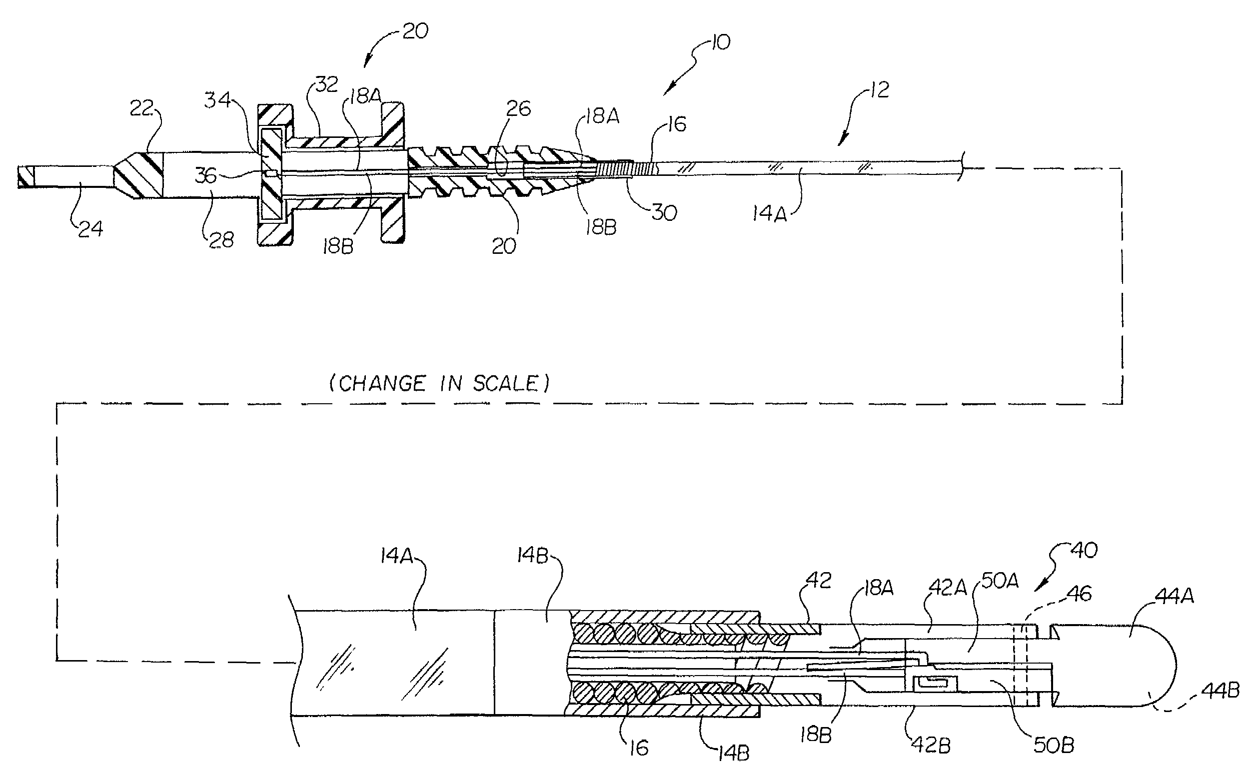
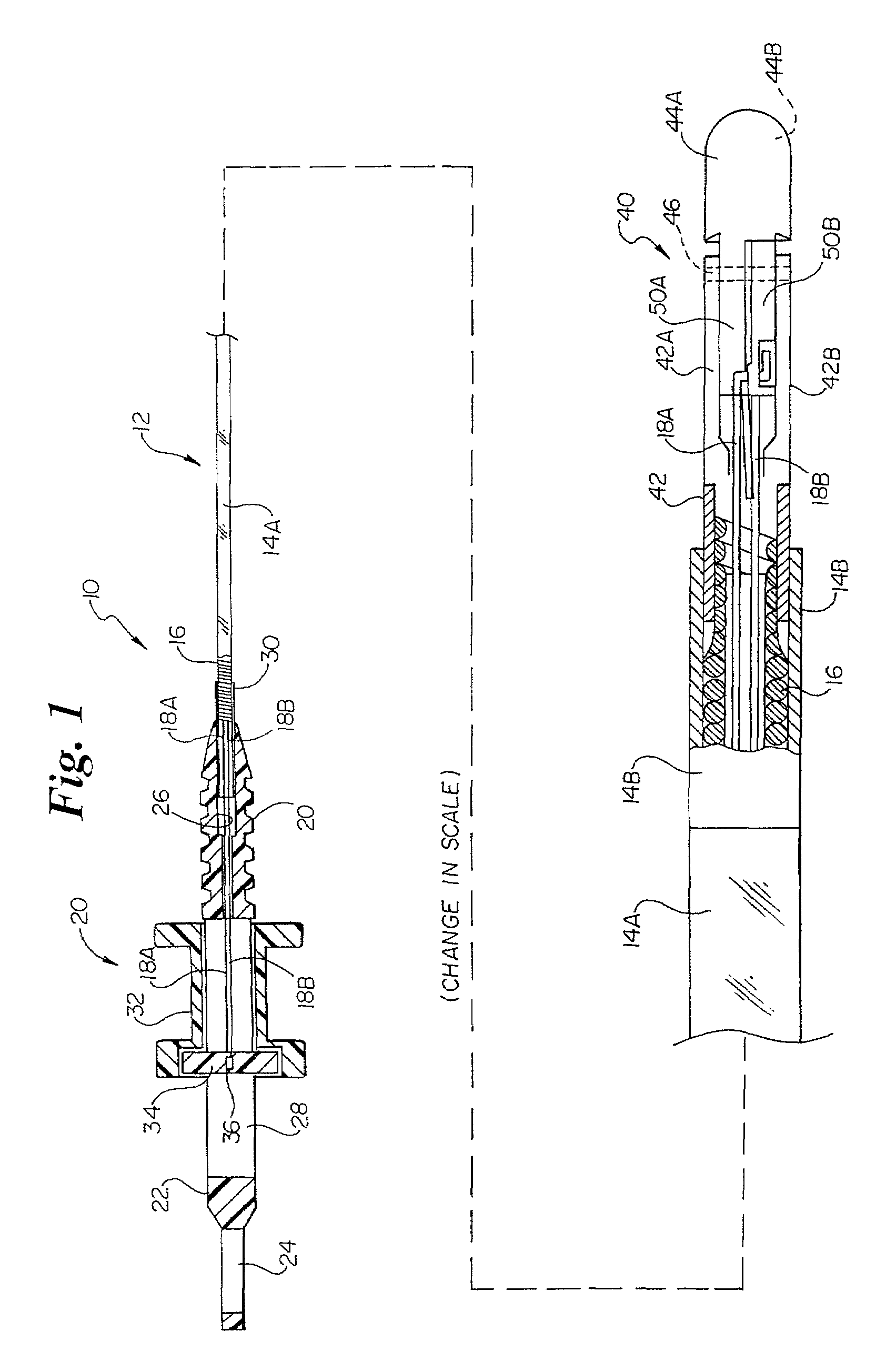
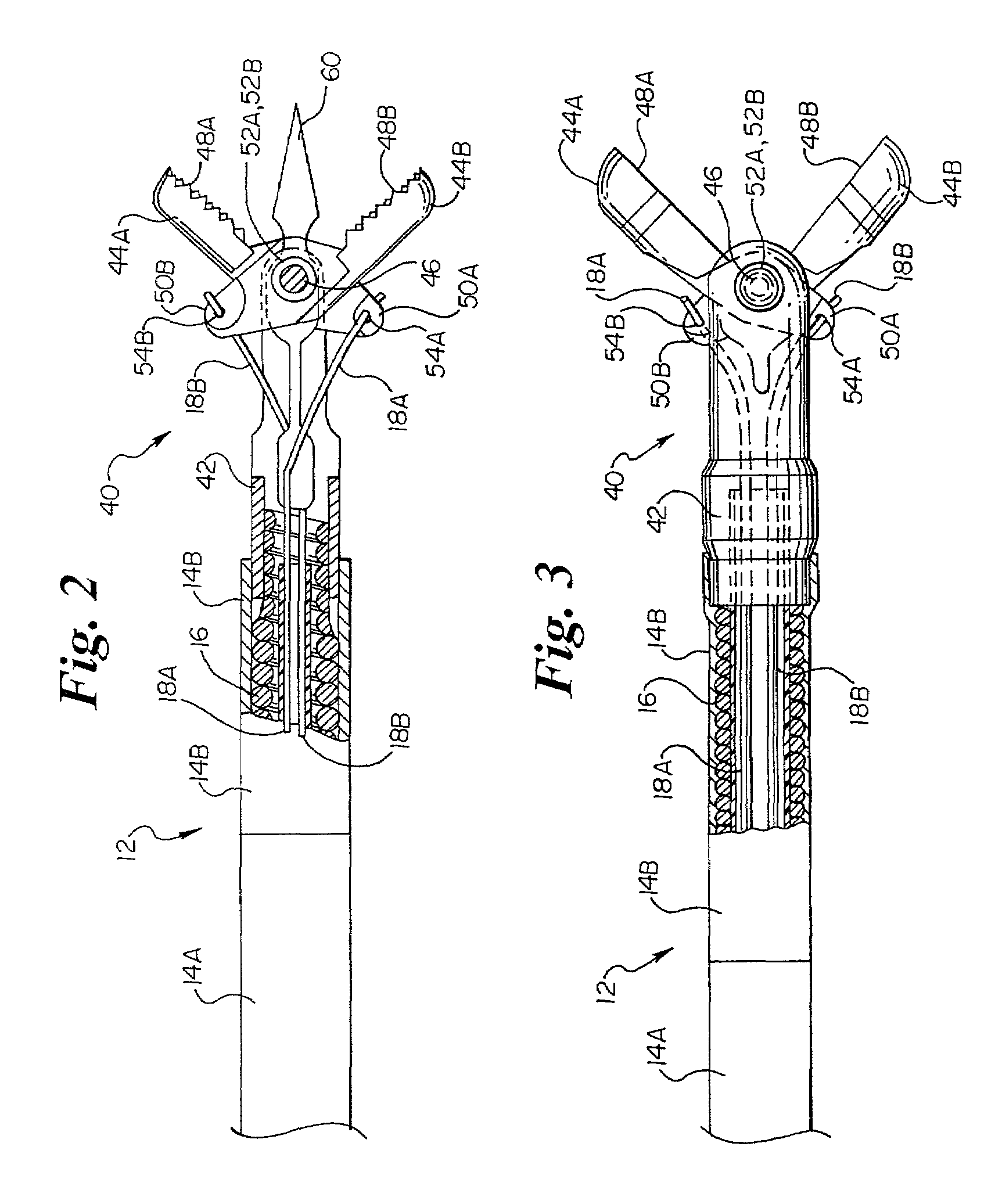
[0011]Refer now to FIG. 1 which illustrates a partially broken elevational view of a biopsy forceps device 10 in accordance with an embodiment of the present invention. The biopsy forceps device 10 includes an elongate shaft 12 having a flexible coil 16 covered by an outer polymeric sheath 14. Polymeric sheath 14 is designed to allow a clinician to easily determine if device 10 had been used previously and / or if device 10 has been adequately cleaned and sterilized.

[0012]The polymeric outer sheath 14 can include a proximal portion 14A and a distal portion 14B. The lengths of the respective portions may be varied in different embodiments. For example, the length of the distal portion 14B may c...


PUM


Abstract

An endoscopic biopsy device having an outer sheath that is transparent, at least along a portion thereof, to allow visualization of at least a portion of the inside of the device. A distal portion of the outer sheath may be relatively more opaque or relatively less reflective to avoid glare that may otherwise result due to the sheath reflecting light from the endoscopic light source.

Description


Claims


Application Information

Patent Timeline
Owner BOSTON SCI SCIMED INC
Learn More