
Method and apparatus for the diagnosis of glaucoma and other visual disorders

a technology for visual disorders and glaucoma, applied in the field of glaucoma and other visual disorders, can solve the problems of slow progression of the disease at typically 1.8% of the field of view per annum, low resolution and inaccurateness, and limiting utility, so as to achieve the effect of detecting the disease in several years, and reducing the number of patients

Inactive Publication Date: 2008-05-06
MCGRATH JOHN ANDREW MURRAY +1
View PDF4 Cites 15 Cited by
  • Summary
  • Abstract
  • Description
  • Claims
  • Application Information

AI Technical Summary

Benefits of technology

This approach significantly enhances the accuracy and speed of visual field measurement, enabling early detection of glaucoma progression and other visual disorders with high reliability and reduced clinical time, making it economically viable for routine screening.

Problems solved by technology

Instruments exist which can measure the field of view of a patient but all available instruments suffer from three major problems that limit their utility in making an early diagnosis.
First they are both low in resolution and inaccurate.
This low resolution means that the slow progression of the disease at typically 1.8% of the field of view per annum can take several years to be detectable.
Secondly the existing devices and methods are slow and complex in clinical use and hence expensive in practitioner time.
This means that even those practitioners who possess a field of view analysis device cannot economically use it as a routine screening device.
Thirdly the existing instruments are inherently expensive and so are not as widely available as is required for the widespread screening necessary for early diagnosis.
At one time it was thought that measurement of eyeball pressure would provide a method for early diagnosis of glaucoma but this has proved unreliable as the correlation between pressure and glaucoma has proved not to be as high as was originally thought.
Instruments for the measurement and mapping of the sensitivity of the human retina known as “visual field analysers” or “Static Auto-perimeters” have hitherto required that the subject perform very unnatural and often uncomfortable eye behaviours such as long periods of attempted fixation on a point.
This process is both slow and prone to inaccuracy.
The ability of the subject to accurately fixate is also known to be poor especially over an extended period and so the accuracy of a purely fixation point to stimulus measurement is further compromised.
Furthermore the disclosed method and apparatus greatly reduces the time required to map the visual field, which makes the test far more economic and practical for routine screening than the existing equipment that requires lengthy tests under expensive expert supervision.
While eye to hand co-ordination and reaction is relatively slow and subject to variability and improvement from practice, and eye to voice reaction time is even slower, the reaction time of the eye itself to stimulus is extremely fast in humans and primates.
Furthermore, because the eye reflex is inherently faster than eye-hand or eye-voice reaction times, any variability in the response has a far lower impact on the accuracy of a reaction dependent measurement.
This vastly reduces the time needed for a diagnostician to establish a subject's field of view.

Method used


Image

  • Method and apparatus for the diagnosis of glaucoma and other visual disorders
  • Method and apparatus for the diagnosis of glaucoma and other visual disorders
  • Method and apparatus for the diagnosis of glaucoma and other visual disorders

Examples


Embodiment Construction

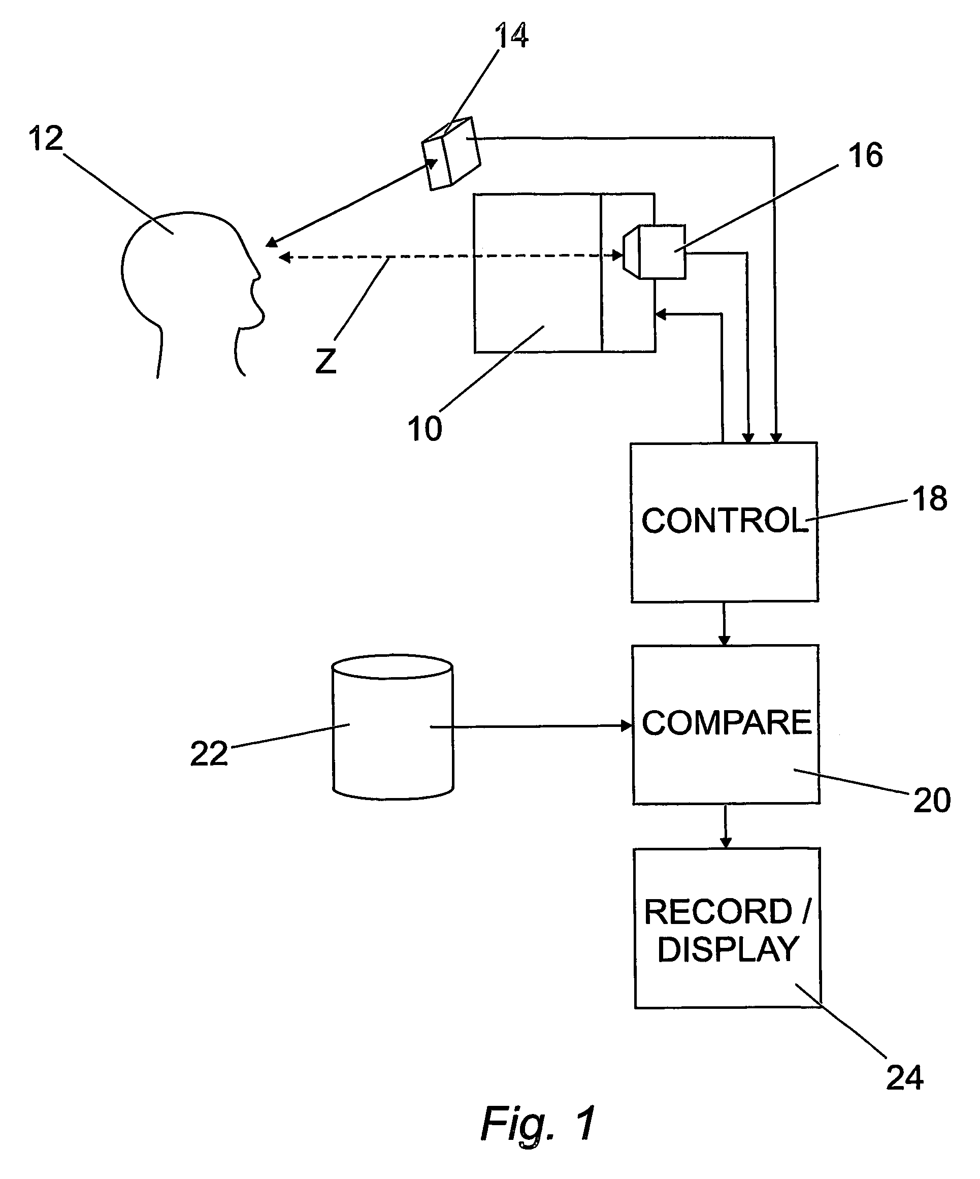
[0016]Embodiments of the invention will be described, by way of example only, with reference to the accompanying drawings, in which:

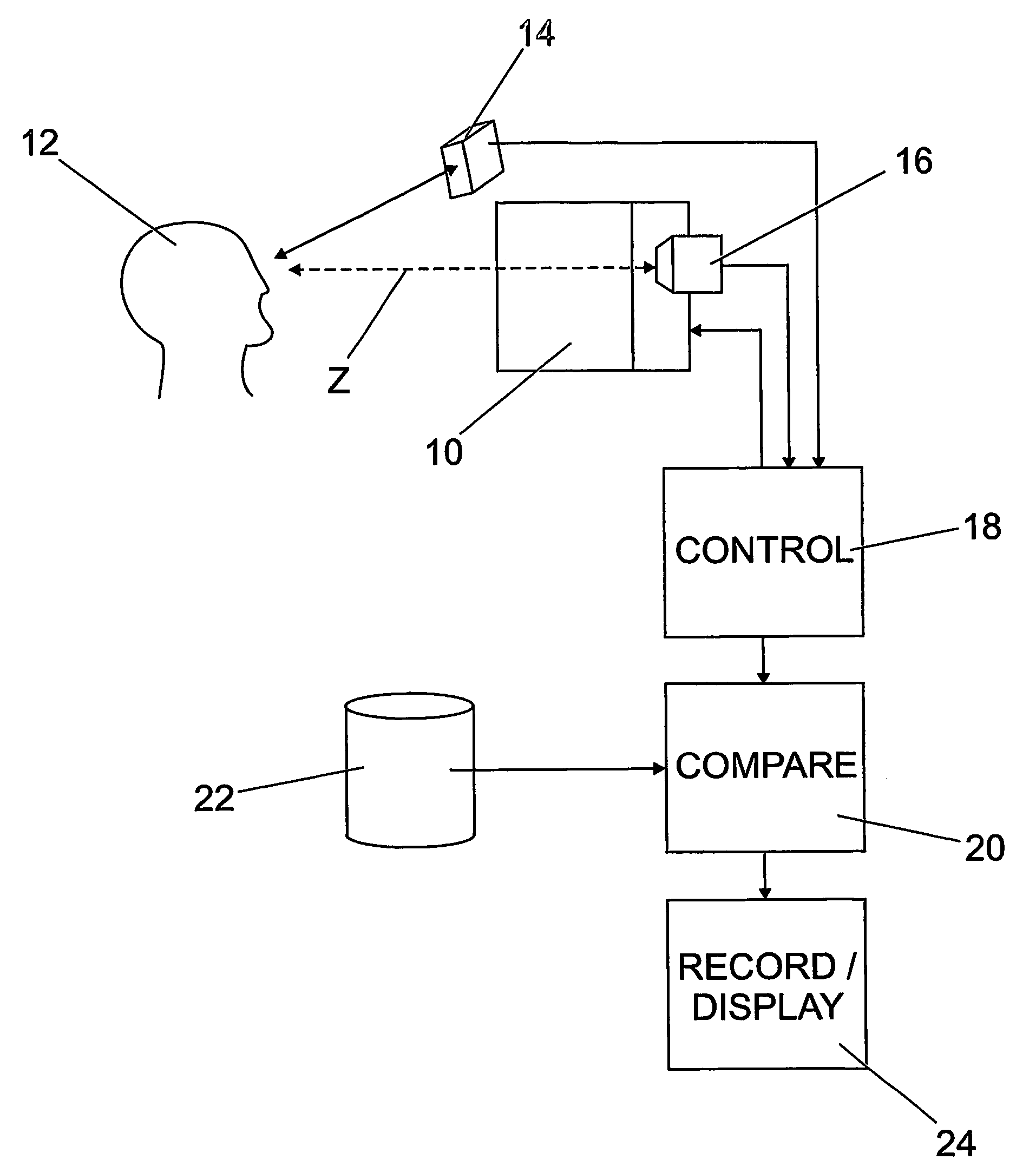
[0017]FIG. 1 is a schematic illustration of one apparatus embodying the invention; and

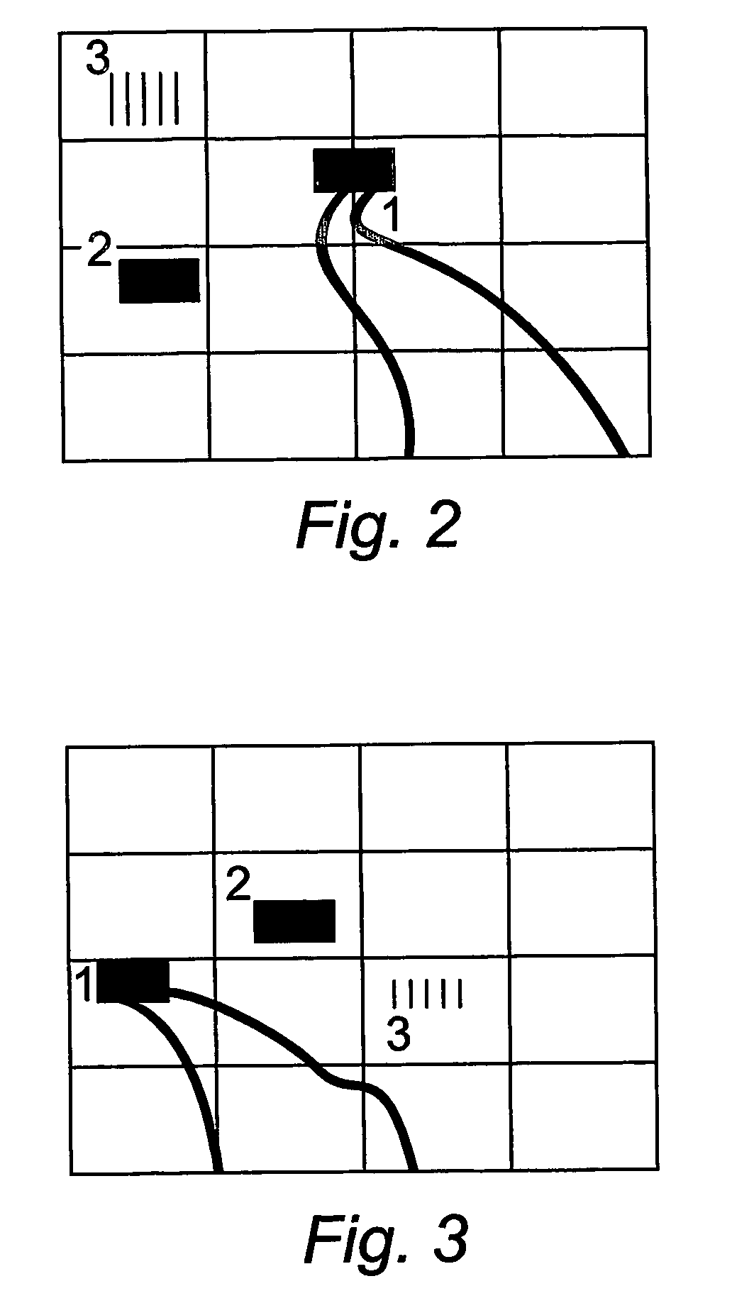
[0018]FIGS. 2 and 3 represent images used in one method according to the invention.

[0019]Prior to this invention visual field analysis methods and apparatus have been extremely crude, in general consisting of an array of lights or other display illuminated under a pseudo-random protocol and at varying brightness straddling the expected threshold of the retina and a fixation point to attempt to maintain some minimal knowledge of the eyes position prior to stimulus. Unfortunately the human vision system is particularly poor at maintaining a constant fixation and furthermore even if this is achieved with practice there are side effects to concentration on a fixation point that significantly reduce the accuracy of the measurement. As a consequence most of these machines ar...


PUM


Abstract

A subject (12) observes an image on a display (10). A control (18) produces a fixation image at a selected position in the display, followed by a stimulus spaced from the fixation image. An eye position sensor (14) detects a saccade movement towards the stimulus. The stimulus is then replaced with a fixation image and the cycle repeated. The time taken to saccade plus the intensity of the stimulus are used to produce a retinal map of field of vision, or to assess other characteristics of the subject.

Description

[0001]This invention relates to methods and apparatus for assessing eye function. The invention is useful inter alia in the diagnosis of glaucoma and other visual disorders, and in the assessment of dyslexia and neurological conditions which affect eye function.[0002]The common form of glaucoma, as is typical of several other visual disorders, is a progressive disease. Currently the disease can be arrested but not cured. Symptoms include the gradual reduction in the field of view of the affected eye progressing in a characteristic pattern. Due to the nature of the human visual system, victims of the disease do not typically notice this reduction in field of view until the disease has already progressed for several years. Instruments exist which can measure the field of view of a patient but all available instruments suffer from three major problems that limit their utility in making an early diagnosis.[0003]First they are both low in resolution and inaccurate. This low resolution me...

Claims


Application Information

Patent Timeline
Patent Type & Authority Patents(United States)
IPC IPC(8): A61B3/14A61B3/024A61B3/032A61B3/113A61B3/12
CPCA61B3/0091A61B3/024A61B3/113A61B3/032A61B3/12
Inventor MCGRATH, JOHN ANDREW MURRAYSTRACHAN, JOHN SCOTT
Owner MCGRATH JOHN ANDREW MURRAY
Learn More