Amelioration of macular degeneration and other ophthalmic diseases

a technology for ophthalmology and macular degeneration, applied in the field of ophthalmological diseases, can solve the problems of oxidative tissue damage, depletion of cellular reactive oxygen species (ros), blood and fluid pooling within the layers of the retina, blurred central vision, etc., to prevent/treat damage to rectal tissue, improve the effect of ophthalmological symptoms, delay or prevent the development of ophthalmological symptoms

Inactive Publication Date: 2005-06-16
COLBY PHARMA CO
View PDF44 Cites 57 Cited by
  • Summary
  • Abstract
  • Description
  • Claims
  • Application Information

AI Technical Summary

Benefits of technology

[0023] According to one aspect of the invention, a method is provided for ameliorating, delaying or preventing the development of, or reducing the symptoms of a disease or disorder of the eye of a patient, wherein the diseases or disorder is macular degeneration, retinopathy glaucoma and uveitis, cystoid macular edema, as well as certain disorders of the cornea, eyelids and conjunctiva, including but not limited to inflammation, trauma, retinal phototoxicity induced by exposure to operating microscope, laser eye injuries, treatment of ablated surface of corneas after excimer laser phototherapy; presbyopia, irradiation damage, corneal neovascularization and various dystrophies such as Fuch's dystrophy, keratoconus, lattice dystrophy and map-dot-fingerprint dystrophy, blepharitis, rosacea of the eye, as well as dry eye syndrome...

Problems solved by technology

As a complex and sensitive organ of the body, the eye can experience numerous diseases and other deleterious conditions that affect its ability to function normally.
Glycosylated proteins generate free radicals, resulting in oxidative tissue damage and depletion of cellular reactive oxygen species (ROS) scavengers, such as glutathione.
The fragile new vessels break easily, causing blood and fluid to pool within the layers of the retina.
In CME, which can occur as a result of disease, injury or surgery, fluid collects within the layers of the macula, causing blurred, distorted central vision.
As it progresses, the traction of the membrane on the macula may cause swelling.
Elevated intraocular pressure (IOP) due to inadequate ocular drainage is a primary cause of glaucoma.
Free radicals and other reactive oxygen species (ROS) cause gradual damage to the trabecular meshwork over a period of time.
Though the IOP does not rise high enough to cause any noticeable symptoms initially, when pressure remains elevated or continues to rise, fibers in the optic nerve are compressed and destroyed, leading to a gradua...

Method used


Image

  • Amelioration of macular degeneration and other ophthalmic diseases
  • Amelioration of macular degeneration and other ophthalmic diseases
  • Amelioration of macular degeneration and other ophthalmic diseases

Examples


example 1

Effect of Tempol-H Treatment on Photochemical Retinal Injury in a Rabbit Model

[0072] The ability of light to produce retinal injury well below the levels capable of producing thermal damage has been termed photochemical retinal injury. The mechanism of action for photochemical retinal injury is believed to be the light induced production of free radicals. The ability of commonly used light sources in clinical ophthalmology to produce photochemical retinal injury is well recognized. The operating microscopes used in ophthalmic surgery have been shown to have the capability of producing retinal photochemical injury. This observation has been utilized to produce a model in the rabbit eye to test the ability of various agents to block photochemical retinal injury. It has been determined that opthalmoscopically visible retinal lesions are detectable after as little as 2.5 minutes of exposure to an operating microscope. The protocol set forth below is utilized to determine whether tempol...

example 2

Evaluation of Tempol-H Incorporation into Retinal Tissue of Rabbits Following Intravitreous Injection

[0075] To assess the efficacy of tempol-H at preventing AMD and other retinal disorders, it must be determined that the drug is incorporated into retinal tissue. The protocol set forth in this example utilizes scintillation technique to establish quantity and duration of tempol-H incorporation into retinal tissue following intravitreous injection in the rabbit. The New Zealand White Rabbit is well characterized in experiments involving intravitreous injection as the large eye of the rabbit provides a suitable template for treatment administration (Hosseini et al., 2003, Lasers Surg. Med. 32: 265-270). Furthermore, the New Zealand White Rabbit has been used extensively in experiments assessing drug uptake into retinal tissue via scintillation technique (Ahmed et al., 1987, J. Pharm. Sci. 76: 583-586).

[0076] Materials and Methods: Twenty-four New Zealand White Rabbits, all male, weig...

example 3

Efficacy Tempol-H in Protection Against Photooxidative Processes in the Retinal Pigment Epithelium (RPE)

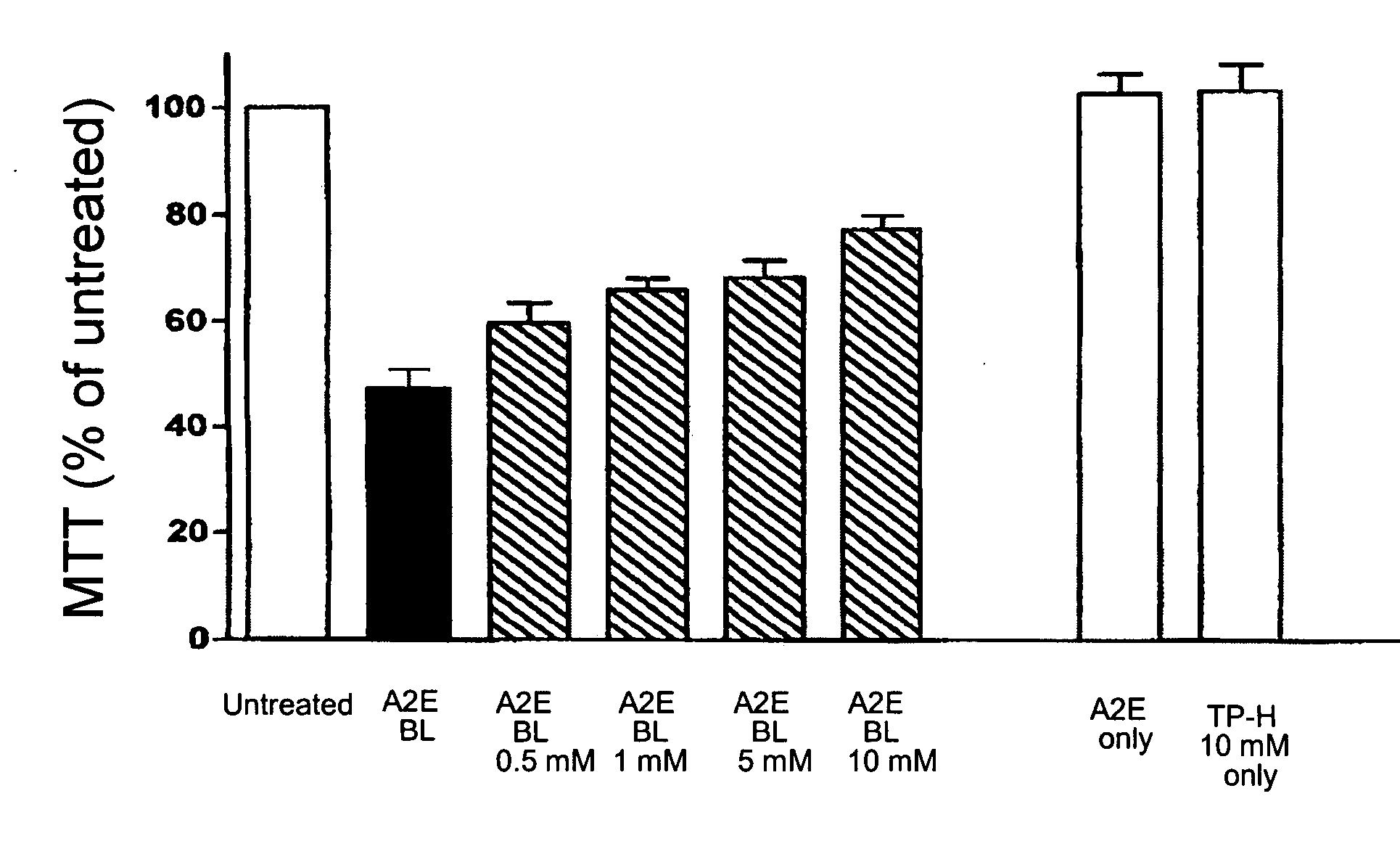
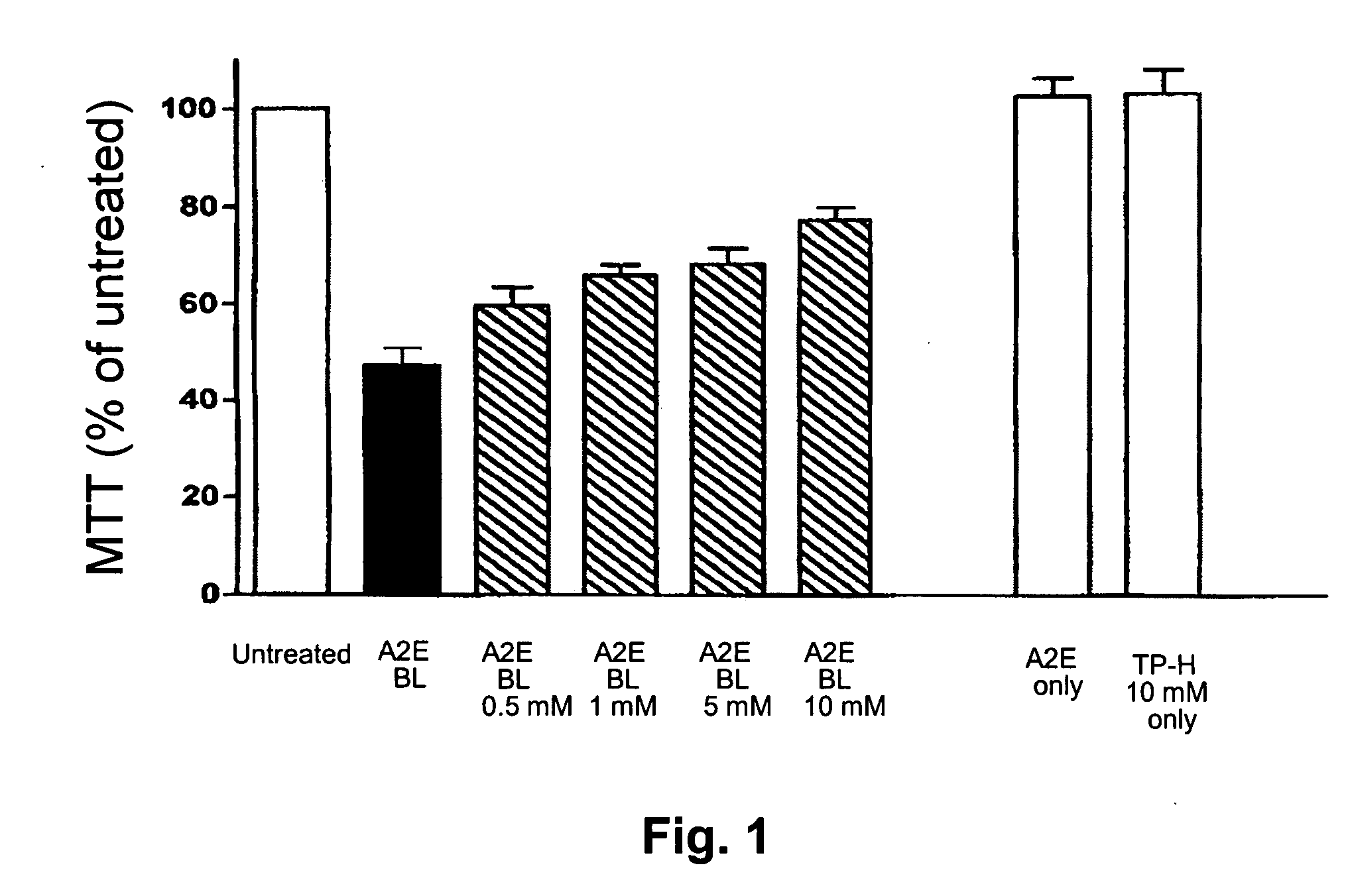
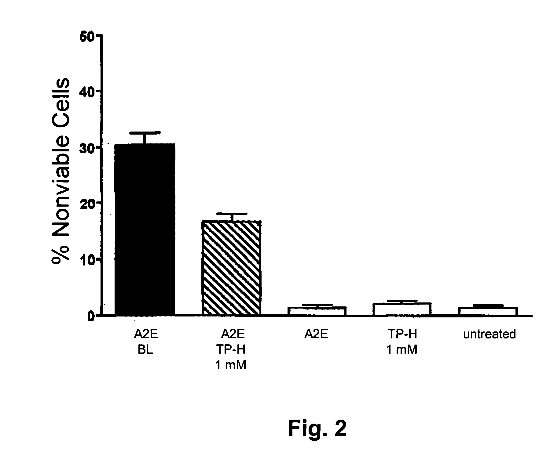
[0082] Background: Although the etiology of atrophic age-related macular degeneration (AMD) is not fully understood, it is generally accepted that AMD begins with the death of retinal pigment epithelial cells, the degeneration of photoreceptor cells, and resultant loss of vision, occurring thereafter. There is a growing body of evidence linking the lipofuscin that accumulates in RPE with the death of these cells. For instance, not only are lipofuscin levels highest in RPE cells underlying the macula, the monitoring of RPE lipofuscin by detection of fundus autofluorescence has also shown that areas of RPE atrophy develop at sites of previously increased fluorescence. Studies concerned with examining associations between RPE lipofuscin and RPE cell death have shown that a major constituent of RPE lipofuscin, the bisretinoid fluorophore A2E, can perturb cellular membranes and can me...


PUM

PropertyMeasurementUnit
Molar densityaaaaaaaaaa

Abstract

Methods for the treatment or prevention of a number of ocular diseases or disorders are disclosed. In particular, methods are disclosed for the amelioration of macular degeneration, diabetic retinopathy and other retinopathies, as well as uveitis, presbyopia, dry eye, glaucoma, blepharitis and rosacea of the eye. The methods comprise administration of a compositions comprising an ophthalmologically acceptable carrier or diluent and a hydoxylamine compound in a therapeutically sufficient amount to prevent, retard the development or reduce the symptoms of one or more of the ophthalmic conditions.

Description

CROSS-REFERENCE TO RELATED APPLICATIONS [0001] This claims benefit of U.S. Provisional Application No. 60 / 523,802, filed Nov. 20, 2003, the entire contents of which are incorporated by reference herein.FIELD OF THE INVENTION [0002] The present invention relates to the treatment of ophthalmic diseases. More specifically, the invention provides methods of reducing or preventing macular degeneration and other deleterious conditions of the retina and surrounding tissues, through the administration of pharmaceutical preparations comprising hydroxylamine compounds. BACKGROUND OF THE INVENTION [0003] Various patents and other publications are referenced herein. The contents of each of these patents and publications are incorporated by reference herein, in their entireties. [0004] As a complex and sensitive organ of the body, the eye can experience numerous diseases and other deleterious conditions that affect its ability to function normally. Many such conditions can be found in the interi...

Claims


Application Information

Patent Timeline
IPC IPC(8): A61KA61K31/13A61K31/198A61K31/445A61K38/05
CPCA61K31/13A61K31/445A61K31/42A61P1/00A61P17/14A61P27/00A61P27/02A61P27/04A61P27/06A61P27/08A61P27/10A61P43/00A61P9/10
Inventor MATIER, WILLIAM L.PATIL, GHANSHYAM
Owner COLBY PHARMA CO
Why Eureka
Try Eureka