

Immunoglobulin fusion proteins
a technology of immunoglobulin and fusion proteins, which is applied in the direction of fusion polypeptides, peptide/protein ingredients, fungi, etc., can solve the problems of heterogeneous product mixtures, high protein-specific factors, and inability to improve the biological activity of a second cytokine, so as to achieve the effect of maximizing the bioactivity of the fused epo
- Summary
- Abstract
- Description
- Claims
- Application Information
AI Technical Summary
Benefits of technology
Problems solved by technology
Method used
Image

Examples
example 1
Construction of Growth Factor / Cytokine-IgG Gene Fusions
A. General Strategy.
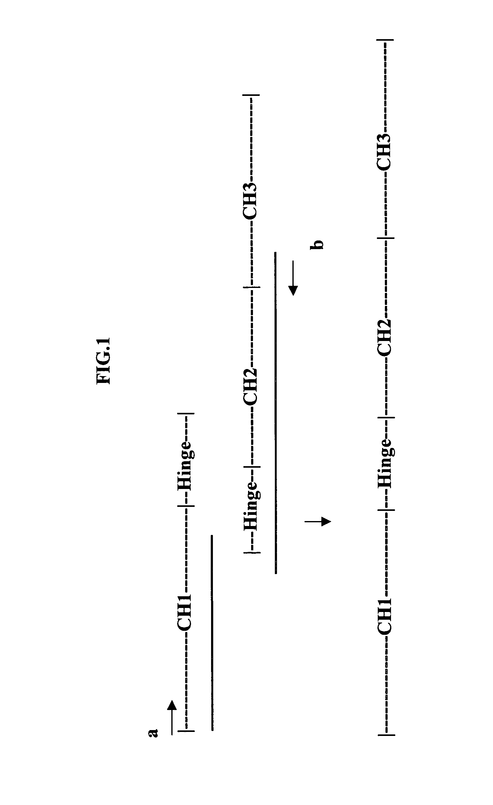
[0047]Growth factor / cytokine (GF)-IgG gene fusions were constructed as described below. The general strategy employed for these constructions is outlined here and the specifics of individual cloning steps are detailed below. Cloning of the IgG4-CH coding sequence involved additional variations to the general strategy and these variations are described below. The human growth factor genes (GH, EPO and G-CSF) were cloned as cDNAs from various RNA sources detailed below. PCR primers used in these clonings added an optimized Kozak sequence (GCCACC; Kozak, 1991) and a Hind III restriction site to the 5′ end of each these clones and a portion of a peptide linker (ser-gly-gly-ser) (SEQ.ID.NO.1) terminating in a Bam HI restriction site, to the 3′ end of each of these clones. The growth factor genes were cloned as Hind III-Bam HI fragments into the mammalian cell expression vector pcDNA3.1 (+) (Invitrogen, Inc., San D...
example 2
Expression and Purification of GF-IgG Fusion Proteins
A. Small Scale Transfection of COS Cells
[0056]Expression and bioactivity of the GF-IgG fusion proteins were assessed initially by small-scale transfection of COS cells. Endotoxin-free plasmid DNAs were prepared using an “Endo-Free Plasmid Purification Kit” (Qiagen, Inc.) according to the vendor protocol and used to transfect COS-1 cells (available from the American Type Culture Collection, Rockville, Md.). The COS-1 cells were grown in Delbecco's Modified Eagle's Media supplemented with 10% FBS, 50 units / ml penicillin, 50 μg / ml streptomycin and 2 mM glutamine (COS cell growth media). Initial transfection experiments were carried out in Costar 6 well tissue culture plates (VWR Scientific) using the following protocol. Briefly, 2-3×10⁵ cells were seeded into each well in 2 ml of COS cell growth media and allowed to incubate overnight at 37° C. and 5% CO2 by which time the cells had reached 50-60% confluency. For each well, 0.8 μg of...
example 3
In Vitro Bioactivities of Purified GF-IgG Fusion Proteins
A. General Strategy
[0063]Cell proliferation assays were developed to measure in vitro bioactivities of the IgG fusion proteins. The assays measure uptake and bioreduction of the tetrazolium salt MTS [3-(4,5-dimethylthiazol-2-yl)-5-3-carboxyphenyl)-2-(4-sulphenyl)-2H-tetrazolium]. In the presence of an electron coupler such as phenazine methosulfate (PMS), MTS is converted to a formazan product that is soluble in tissue culture media and can be measured directly at 490 nm. Cell number is linear with absorbance values up to about 2. For EPO and G-CSF we were able to use existing cell lines to develop the bioassays. For GH, we needed to create a cell line that proliferates in response to GH. Such a cell line was created by stably transforming a murine leukemia cell line with a rabbit GH receptor.
[0064]In general, the bioassays were set up by washing the appropriate cells three times with media (no additives) and resuspending the ...
PUM
| Property | Measurement | Unit |
|---|---|---|
| Molar density | aaaaa | aaaaa |
| Molar density | aaaaa | aaaaa |
| Molar density | aaaaa | aaaaa |
Abstract
Description
Claims
Application Information