X-ray CT apparatus

a ct and x-ray technology, applied in the field of x-ray ct (computed tomography) apparatus, can solve the problems of large x-ray needless exposure problem, hard control of image quality, etc., and achieve the effect of reducing exposur

Inactive Publication Date: 2010-08-17
GE MEDICAL SYST GLOBAL TECH CO LLC
View PDF14 Cites 19 Cited by
  • Summary
  • Abstract
  • Description
  • Claims
  • Application Information

AI Technical Summary

Benefits of technology

[0006]Therefore, an object of the present invention is to provide an X-ray CT apparatus capable of obtaining each tomographic image indicative of X-ray tube voltage dependent information of a subject with the optimum image quality by less reduced exposure.
[0020]According to the X-ray CT apparatus of the present invention, an advantageous effect is brought about in that an X-ray CT apparatus capable of obtaining each tomographic image indicative of X-ray tube voltage dependent information of a subject with the optimum image quality by more reduced exposure can be realized.

Problems solved by technology

However, the process of bringing the information about the difference between the dependencies of the X-ray absorption coefficients on the X-ray tube voltages was a process hard to control the quality of each image obtained by a process for performing weighted subtraction on the tomographic images at the X-ray tube voltages and control noise of the image.
On the other hand, the X-ray CT apparatus having the multi-row X-ray detector or the X-ray CT apparatus based on the two-dimensional X-ray area detector typified by the flat panel has a tendency that as a cone angle of an X-ray cone beam becomes great, the problem of X-ray needless exposure becomes larger.

Method used


Image

  • X-ray CT apparatus
  • X-ray CT apparatus
  • X-ray CT apparatus

Examples


Embodiment Construction

[0047]The present invention will hereinafter be described in further detail by embodiments illustrated in the drawings. Incidentally, the present invention is not limited thereby.

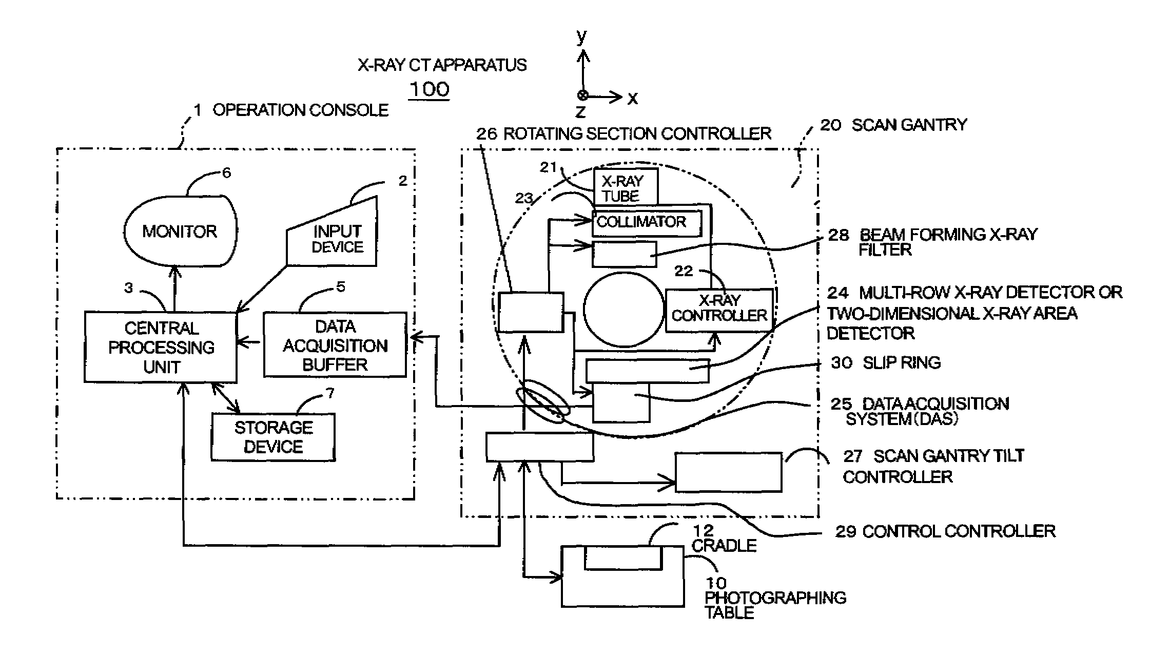
[0048]FIG. 1 is a configuration block diagram of an X-ray CT apparatus according to one embodiment of the present invention. The X-ray CT apparatus 100 is equipped with an operation console 1, a photographing table 10 and a scan gantry 20.

[0049]The operation console 1 is equipped with an input device 2 which receives an input from an operator, a central processing unit 3 which executes a pre-process, an image reconstructing process, a post-process, etc. a data acquisition buffer 5 which acquires or collects X-ray detector data acquired by the scan gantry 20, a monitor 6 which displays a tomographic image image-reconstructed from projection data obtained by pre-processing the X-ray detector data, and a storage device 7 which stores programs, X-ray detector data, projection data and X-ray tomographic images t...


PUM


Abstract

The present invention provides an X-ray CT apparatus capable of obtaining each tomographic image indicative of X-ray tube voltage dependent information of a subject with the optimum image quality by less reduced exposure. The X-ray CT apparatus includes device for setting a plurality of X-ray tube voltages and sets imaging conditions used in the photography using the respective X-ray tube voltages in such a manner that respective image noise of tomographic images photographed at the X-ray tube voltages become substantially identical to one another. The setting of the imaging conditions is the setting of X-ray tube currents. The X-ray tube currents are set based on geometrical characteristic amounts of the subject determined from each scout image in such a manner that the respective image noise of the tomographic images photographed at the plurality of X-ray tube voltages become substantially identical to one another.

Description

CROSS REFERENCE TO RELATED APPLICATIONS[0001]This application claims the benefit of Japanese Patent Application No. 2007-290817 filed Nov. 8, 2007, which is hereby incorporated by reference in its entiretyBACKGROUND OF THE INVENTION[0002]The subject matter disclosed herein relates to a technique of a medical X-ray CT (Computed Tomography) apparatus or an industrial X-ray CT apparatus.[0003]In an X-ray CT apparatus having a multi-row X-ray detector or an X-ray CT apparatus based on a two-dimensional X-ray area detector of a matrix structure typified by a flat panel, there has heretofore been a technique which photographs tomographic images at a plurality of X-ray tube voltages as shown in FIG. 7 and brings information about a difference between the dependencies of X-ray absorption coefficients of materials constituting a subject on the X-ray tube voltages into imaging, based on the tomographic images.BRIEF DESCRIPTION OF THE INVENTION[0004]However, the process of bringing the informa...

Claims


Application Information

Patent Timeline
Patent Type & Authority Patents(United States)
IPC IPC(8): A61B6/00
CPCA61B6/032A61B6/4085A61B6/466A61B6/488A61B6/542
Inventor NISHIDE, AKIHIKOSASAKI, KOSUKEHORIUCHI, TETSUYA
Owner GE MEDICAL SYST GLOBAL TECH CO LLC
Why Eureka
Try Eureka