Cannula insertion device and related methods

a technology of cannula and insertion device, which is applied in the field of systems and methods for inserting cannulas into patients, can solve the problems of obstructing the attached tube and medical device, affecting the patient's comfort,

Active Publication Date: 2010-12-07
CEQUR SA
View PDF82 Cites 170 Cited by
  • Summary
  • Abstract
  • Description
  • Claims
  • Application Information

AI Technical Summary

Benefits of technology

[0007]During an insertion procedure described herein, a cannula is used in conjunction with a needle. The needle is used to penetrate the body. Subsequently, the needle is withdrawn from the body, allowing the cannula to remain in its inserted position. Consequently, one advantage of the invention is to increase the comfort of a patient during the insertion and withdrawal of a catheter or cannula. This benefit may be achieved with a device for the insertion of a cannula of the type described herein, wherein the needle, after reaching its extended state, can be withdrawn quickly and automatically by a retraction apparatus.
[0008]One embodiment of the invention allows the needle to not only quickly penetrate the body tissue (i.e., blood vessel, organ, or other body part) of a patient, but due to aid from the retraction apparatus, the needle is quickly withdrawn from the body tissue and simultaneously, the inserted cannula remains within the body tissue. In this way, since the needle is relatively quickly withdrawn from the body tissue, a much more comfortable sensation is experienced than that associated with a slower insertion and withdrawal. Further, any sensation is transient. The retraction apparatus is activated, in any case, when the needle is already within the patient's tissue, that is to say, when the needle is in its extended position. Thereby, it is possible for the operator to insert the needle and its associated cannula to a sufficient depth to reach a desired position within the body tissue.
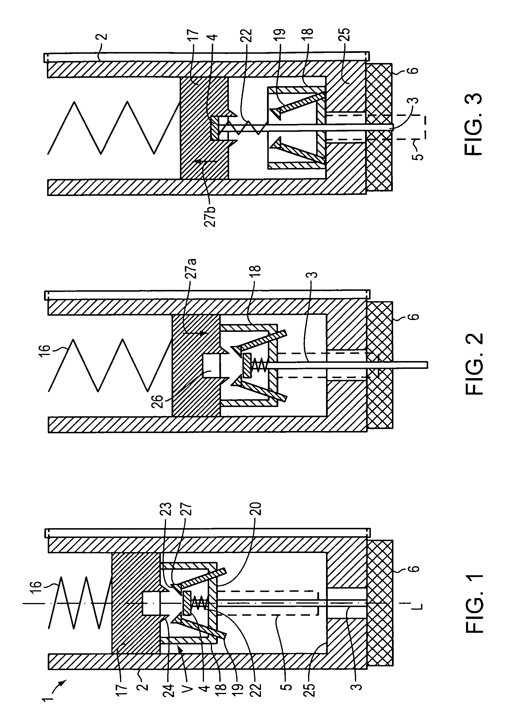
[0009]In one embodiment, the driving element or needle plunger is subject to the force of a spring. The spring can be prestressed and restrained by an arresting apparatus from which it is released to carry out its driving function. The spring acts upon a drive mechanism, which includes at least one first part (e.g., a plunger), which is in driving contact with a free end of the spring and the drive mechanism includes one separate second part (e.g., a support structure), indirectly activated by the spring. Thus, the second part is made slidingly displaceable in a first direction by means of the motion of the first part and, conversely, separates itself therefrom upon a movement of the first part in an opposite direction. Such an arrangement reduces the weight, which contributes to momentum during the retraction of the needle. Simultaneously, the needle, upon its movement from the retracted position into the extended position, stabilizes the cannula sufficiently when so projected, so that that the insertion procedure can be reliably carried out.
[0010]In certain embodiments, the needle, which is seated in the second part, is releasably held. Accordingly, the needle can disengage itself from the second part at the outset of a retracting motion and can be withdrawn without obstruction. In this way, the needle is not permanently affixed to the drive mechanism, an advantage which can be usefully employed to even move the needle more quickly, i.e., through more rapid increments of retractive motion.
[0011]At the time that the needle is disconnected from the second part, nothing more than the weight of the needle need be placed in motion, an advantage which allows undiminished acceleration. Since the movement of a needle within the body tissue can be uncomfortable, it is helpful if the displacement of the needle can be accelerated so that it can be removed from the tissue in a minimal amount of time.
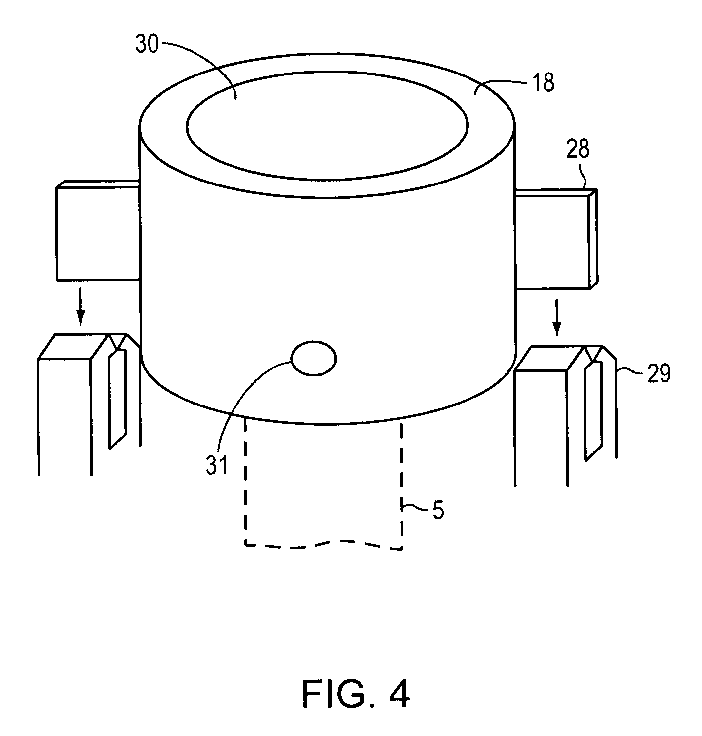
[0012]In one embodiment, the activation of the needle is subjected to force by a separate retraction spring (compression type) dedicated thereto, which acts in a direction from the second part toward the first part. It is possible, then, that this retraction spring serving the needle can be employed to force the now-released needle away from the second part in a direction that withdraws it from the penetrated body tissue. The second part can include at least one holding element that retains the needle therein, and permits the needle to be subsequently retracted from an extended position. The concept of “retraction” or “withdrawal,” as used herein, is used to describe removal of the needle from the lumen of the cannula after full insertion or extension of the cannula. The method of construction of the holding element, that is, as to whether its retaining characteristic is due to mechanical, magnetic, or other induced forces, is of secondary importance for the purpose of this description. During the interim within which the needle has not yet reached its extended position, the holding element acts to affix the needle within the second part. As soon as the second part has reached its final and extended position, the retention of the needle is no longer necessary. Accordingly, at this time, the holding element releases the needle, so that it departs from the second part and extricates itself therefrom.

Problems solved by technology

When performed manually, the penetration of the catheter-carrying needle into the body, however, may be considered uncomfortable for a patient, or may cause some other anxiety.
Also, the subsequent manual withdrawal of the needle is often regarded by some patients as painful.
The above-described insertion sets may be acceptable for some patients, but the attached tube and medical device can be obtrusive, which may be undesirable in some cases.

Method used


Image

  • Cannula insertion device and related methods
  • Cannula insertion device and related methods
  • Cannula insertion device and related methods

Examples


Embodiment Construction

[0048]FIG. 1 depicts an embodiment of an insertion device for a catheter 1 having a housing 2, within which a needle 3 is affixed by a needle holder 4. On the end remote from the needle holder 4, the needle 3 carries a catheter or cannula 5. In this document, the terms “catheter” and “cannula” are used interchangeably and without limitation to described any tube-like implement to deliver fluids to any body part of a patient. In the attached figures, for the sake of clarity, the representation of the catheter 5 is enlarged. In general, the diameter of the catheter 5 is only slightly larger than the outside diameter of the needle 3.

[0049]The needle 3, in a manner not shown, is so enveloped by a guide in the housing 2, that it can only move in a direction parallel to its longitudinal axis L. The design allows the needle 3 to be displaced into an extended position. In this extended position of the needle 3, depicted in FIG. 2, the catheter 5 is positioned at least partially outside the ...


PUM


Abstract

A cannula insertion device includes a housing defining an opening for receiving therethrough a cannula and further defining a channel, and a cannula forming a lumen, the cannula adapted for sliding movement within the housing from a retracted position to an extended position. When the cannula is in the retracted position, the lumen is located remotely from the channel and the channel is in fluidic communication with the opening. When the cannula is in the extended position, the lumen is in fluidic communication with the channel.

Description

CROSS-REFERENCE TO RELATED APPLICATION[0001]This application claims priority to and the benefit of German Patent Application Ser. No. 10 2007 049 446.9, filed Oct. 16, 2007, the disclosure of which is hereby incorporated by reference herein in its entirety.FIELD OF THE INVENTION[0002]This invention relates generally to systems and methods for inserting a cannula into a patient and, more specifically, to a device that automatically inserts a cannula, as well as connects the cannula lumen to a channel within an infusion device.BACKGROUND[0003]In many cases, it is necessary to insert a catheter or cannula into the body, or a body part of, a patient. By means of the so-inserted cannula, it is then possible to infuse medicinally-required fluid or material. Another reason for the employment of a cannula may be to aid in insertion of a micro-dialysis probe into the body.[0004]To insert a cannula into a patient, a rigid needle is placed within the lumen of the cannula, and the needle and ca...

Claims


Application Information

Patent Timeline
Patent Type & Authority Patents(United States)
IPC IPC(8): A61M5/178
CPCA61M5/14248A61M5/158A61M25/02A61M25/0606A61M2005/14252A61M2005/1583A61M2005/1585A61M5/3287
Inventor GRAVESEN, PETERARNDT, HEIKO
Owner CEQUR SA
Why Eureka
Try Eureka