
Method of removing antibody free light chains from blood

a free light chain and antibody technology, applied in the field of blood free light chain removal, can solve the problems of more entering the distal tubule, urine concentration of flc not being considered to accurately reflect plasma cell synthesis, and high sensitivity of the technique, so as to reduce the circulating il-6 level, reduce the loss of albumin, and restore the immune responsiveness of blood cells

Active Publication Date: 2011-01-25
GAMBRO LUNDIA AB +1
View PDF5 Cites 47 Cited by
  • Summary
  • Abstract
  • Description
  • Claims
  • Application Information

AI Technical Summary

Benefits of technology

[0023]The applicants postulated that removal of serum free light chains by haemodialysis might be a way of reducing renal failure in patients with multiple myeloma (Bradwell et al. (2005) Blood 106 (11) 3482: 972a; 47th Annual Meeting of American Society of Hematology, 10-13 Dec. 2005). The applicants have surprisingly found that a patient having excess serum free light chains can be treated by dialysis, with the result that free light chain concentration is reduced.
[0026]Morgera, et al. show a remarkable clearance of interleukin-6 with high cut-off treatments leading to a significant decrease in circulating IL-6 levels in septic patients having acute renal failure (Morgera, et al. Intensive Care Med. (2003), 29: 1989-1995). Furthermore, the treatment lead to a restoration of immuneresponsiveness of blood cells in those patients (Morgera S., et al., Nephrol. Dial. Transplant (2003), 18: 2570-2576). A study where patients were randomly allocated to high cut-off CVVH or high cut-off CVVHD showed that convention and diffusion did not exhibit the expected difference in terms of clearance of middle-molecular-weight proteins, whereas using diffusion instead of convention significantly reduces the loss of albumin while maintaining good cytokine clearance rates. In CVVHD mode a maximum albumin loss of 950 mg per hour in patients treated with the HCO membrane was reported (Morgera S., et al. Am. J. Kidney Dis. (2004), 43: 444-453). The applicants have now surprisingly found that such membranes can be used to effectively reduce sFLC concentrations.

Problems solved by technology

Furthermore, the sensitivity of the technique is very high.
Historically, urine concentrations of FLC have not been considered to accurately reflect plasma cell synthesis.
Subsequently, and with increasing renal failure, hyperfiltering glomeruli leak albumin and other proteins which compete with FLCs for absorption, thereby causing more to enter the distal tubules.
The casts obstruct tubular fluid flow, leading to disruption of the basement membrane and interstitial damage.
Rising concentrations of sFLCs are filtered by the remaining functioning nephrons leading to a vicious cycle of accelerating renal damage with further increases in sFLCs.
Furthermore, highly polymerised FLCs (a frequent finding in MM) are probably not nephrotoxic because they cannot readily pass through the glomeruli.
Since the normal daily production of FLC is ˜500 mg, increases in ˜5 g / day are likely to be nephrotoxic in many patients.
However, FLC excretion is an indicator of renal damage in addition to its cause.

Method used


Image

  • Method of removing antibody free light chains from blood
  • Method of removing antibody free light chains from blood
  • Method of removing antibody free light chains from blood

Examples


examples

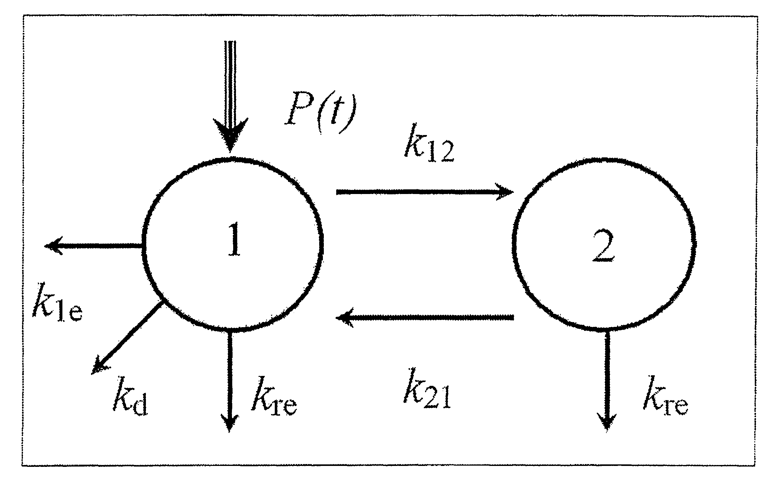
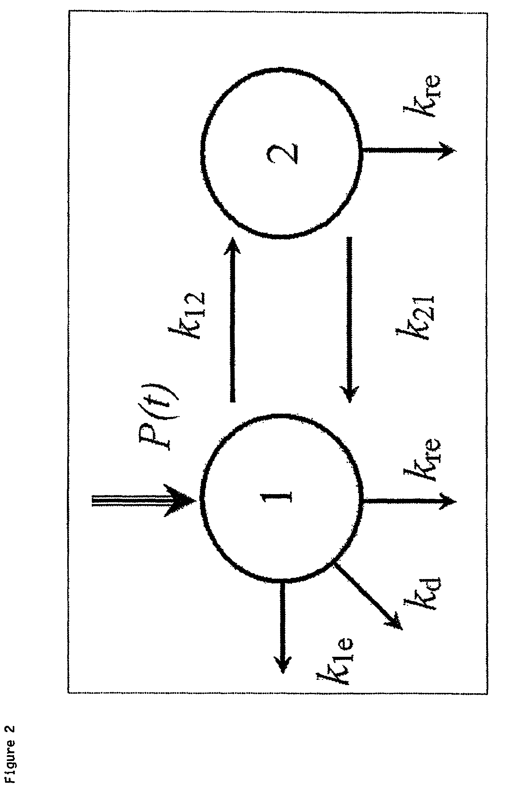
[0076]Embodiments of the invention will now be shown, by way of example only, with reference to Tables, 1-5, and FIGS. 2-8. Serum and dialysate concentrations of free light chains in the examples below were determined by standard free light chain assays available under the trademark “Freelite” from The Binding Site Ltd., Birmingham, UK. However, other methods of detecting the concentration of free light chains, which are known in the art, could also have been used.

Patients and Methods

[0077]This study was approved by the Solihull and South Birmingham Research Ethics Committees and the Research and Development Department of the University Hospitals Birmingham NHS Foundation Trust. All patients gave informed and written consent.

Study Design and Participants

[0078]The study comprised:—1) An initial in-vitro and in-vivo assessment of dialysers for clearance of FLCs, 2) Development of a compartmental model for FLC removal based upon observed dialysis results and 3) Use of the model and the...


PUM

PropertyMeasurementUnit
molecular weightaaaaaaaaaa
diameteraaaaaaaaaa

Abstract

There is provided a method of reducing blood free light chain concentration in a subject, the method comprising the step of subjecting the subject's blood to haemodialysis, haemodiafiltration or haemofiltration. There is also provided the use of a dialysis membrane which is a membrane that allows passage of molecules having a molecular weight of up to 45 kDa in presence of whole blood, and has a molecular weight exclusion limit in water of about 200 kDa in the preparation of a haemodialysis unit for conducting haemodialysis, haemodiafiltration or haemofiltration on a subject to reduce blood free light chain concentration in the patient.

Description

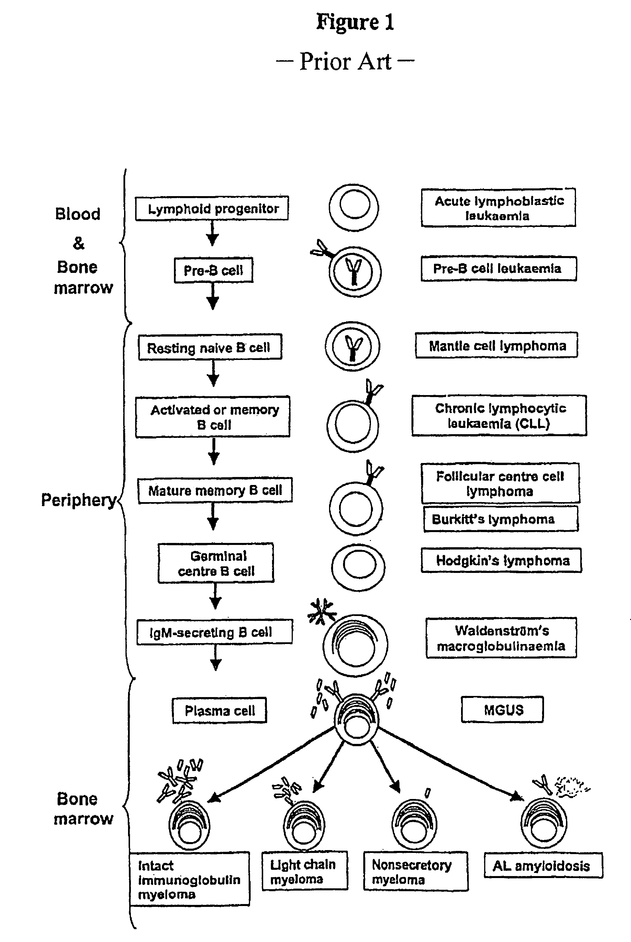
CROSS REFERENCE TO RELATED APPLICATION[0001]This application claims the benefit under 35 U.S.C. §119(a) of GB Application Serial No. 0608444.6, filed Apr. 27, 2006, the disclosure of which is incorporated herein by reference.TECHNICAL FIELD[0002]The present invention relates to the field of removing free light chains from blood, particularly methods for such removal and apparatus for use in such methods.BACKGROUND OF THE INVENTION[0003]Antibody molecules (also known as immunoglobulins) have a twofold symmetry and are composed of two identical heavy chains and two identical light chains, each containing variable and constant domains. The variable domains of the heavy and light chains combine to form an antigen-binding site, so that both chains contribute to the antigen-binding specificity of the antibody molecule. The basic tetrameric structure of antibodies comprises two heavy chains covalently linked by a disulphide bond. Each heavy chain is in turn attached to a light chain, again...

Claims


Application Information

Patent Timeline
Patent Type & Authority Patents(United States)
IPC IPC(8): B01D11/00B01D61/00C02F1/44A61M1/34A61M1/16
CPCA61M1/16A61M1/34B01D69/08B01D69/12B01D2325/02A61M1/1613A61M2205/3334A61M1/3406
Inventor BRADWELL, ARTHUR RANDELLGOEHL, HERMANNSTORR, MARKUS
Owner GAMBRO LUNDIA AB
Learn More