Method for assisting with percutaneous interventions

a percutaneous intervention and percutaneous diagnosis technology, applied in the field of percutaneous intervention assistance, can solve the problems of a serious puncture error, a risk of dying from such an intervention, and some limitations of percutaneous diagnosis and therapy, so as to simplify the workflow, facilitate the registration of imaging systems, and improve the accuracy of puncture errors

Active Publication Date: 2012-01-17
SIEMENS HEALTHCARE GMBH
View PDF5 Cites 6 Cited by
  • Summary
  • Abstract
  • Description
  • Claims
  • Application Information

AI Technical Summary

Benefits of technology

[0008]The object of the present invention is to specify a method for assisting with percutaneous interventions, which is suitable for monitoring biopsies and punctures and which reduces the radiation load for the patient during the intervention.
[0011]With the proposed method ultrasound imaging with an external ultrasound device is thus combined with 3D x-ray imaging using a C-arm x-ray system or a robot-based x-ray system. An external ultrasound system here refers to an ultrasound system, which is arranged outside the body and is therefore not inserted into the body. A robot-based x-ray system has an imaging system guided by one or more robot arms. With the robot-based x-ray system 2D x-ray images of the object region can be recorded at different projection angles in the same manner as with an x-ray C-arm system, these then being used to reconstruct a 3D x-ray image data record. Ultrasound imaging during the intervention has the advantage of imaging in real time without a radiation load for the patient. On the other hand high resolution is achieved with the x-ray imaging carried out before the intervention. The combination of the C-arm or robot-based x-ray system with external ultrasound imaging has the advantage that in many instances registration can be simpler than when using a CT system, which generally requires the patient to be moved from room to room for the CT recording and the intervention. The C-arm x-ray system or robot-based x-ray system in contrast provides very easy access to the patient and can therefore be deployed in the same room, in which the intervention also takes place. Spatial registration of the ultrasound system and x-ray system is therefore simplified significantly.
[0020]The proposed method allows simpler and more precise registration of the imaging systems and as a result greater puncture accuracy to be achieved. All modalities are accessible in one room, without having to move the patient from room to room. This simplifies the workflow. Ultrasound imaging during the intervention minimizes the ionizing radiation load on the patient. The 3D x-ray image data record created before the intervention can also be used to plan the intervention, so there is no need for the previously standard creation of a CT image data record for pre-intervention planning. The merging of x-ray image data and ultrasound image data increases the advantages of both imaging modalities.

Problems solved by technology

Nevertheless percutaneous diagnosis and therapy still have some limitations.
However there is still a risk of dying from such an intervention, in particular if a major vessel has been damaged accidentally.
Puncture error is a further serious problem.
Puncture errors can result in false negative diagnoses or an inadequate therapeutic dosage in the required target region.
The problem with a precise puncture lies in the variability of the target region due to respiratory movement, displacement and deformation of organs and the disease center due to the advance of the needle and possible unpredictable patient movement.
However with the last-mentioned method either the deposition of ionizing radiation during the intervention must be taken into account for the patient or the application is limited to certain regions in the case of the ultrasound catheter.
In particular biopsies and punctures can generally not be carried out using an ultrasound catheter.

Method used


Image

  • Method for assisting with percutaneous interventions
  • Method for assisting with percutaneous interventions

Examples


Embodiment Construction

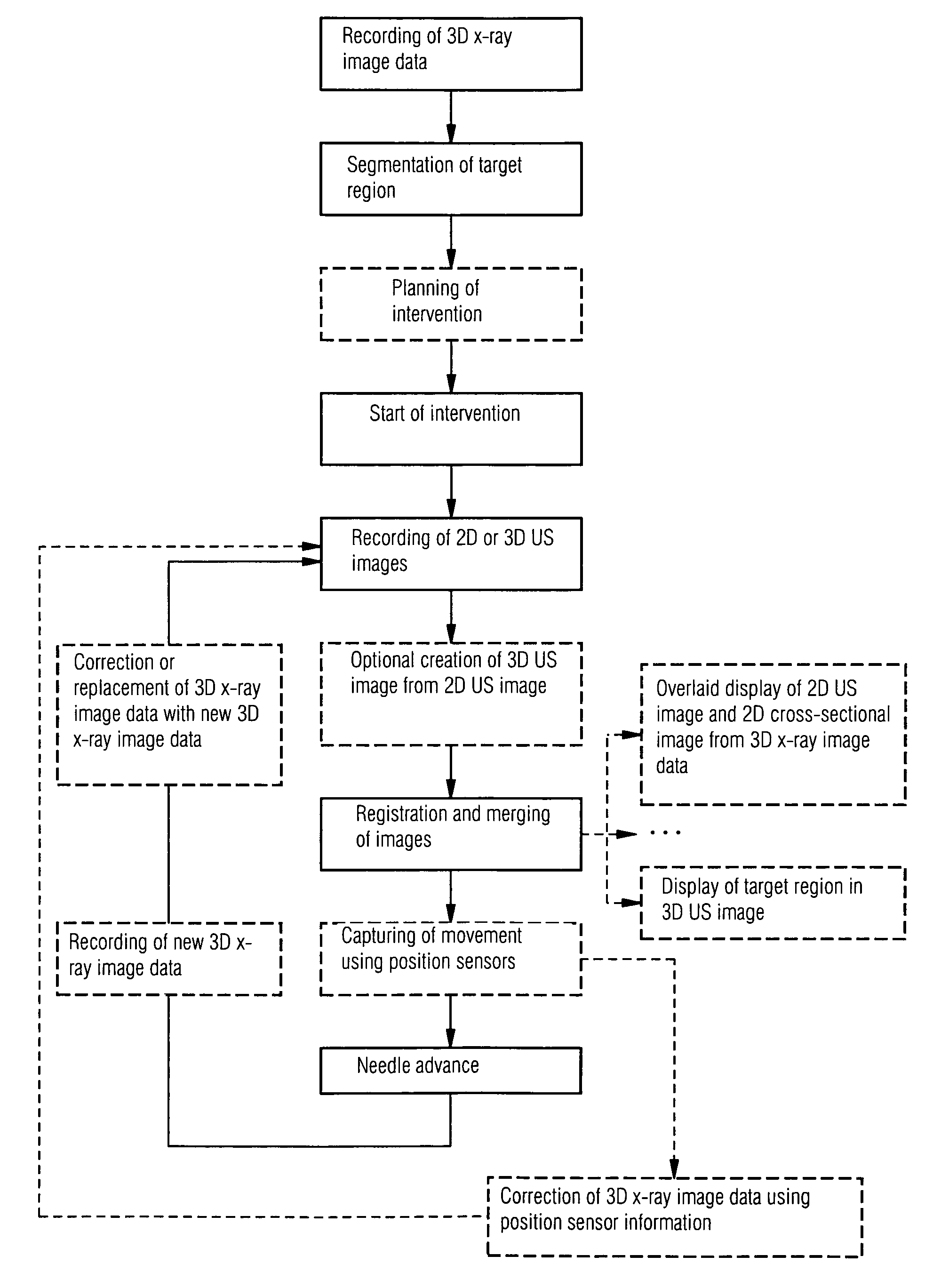
[0022]The workflow of the proposed method is described here with reference to a liver biopsy. The workflow comprises the following steps:[0023]Step 1: Recording and reconstruction of a 3D DynaCT data record of the liver with high resolution; segmentation of the target region, for example a tumor, and possibly color display in the overall DynaCT data record.[0024]Step 2: Recording of a 3D ultrasound image using an appropriate ultrasound head for 3D imaging or a number of 2D ultrasound images for a known spatial position of the ultrasound head; optionally segmentation of the target region in said ultrasound images.[0025]Step 3: Registration of the recorded ultrasound images (2D or 3D) with the DynaCT data record.[0026]Step 4: Needle advance with image monitoring until the needle has reached the target region in the liver.

[0027]The color display of the tumor in step 1, which can be an option, serves to enhance the identifiability of the tumor for the user in the respective image displa...


PUM


Abstract

The present invention relates to a method for assisting with percutaneous interventions, wherein 2D x-ray images of an object region are recorded before the intervention using a C-arm x-ray system or a robot-based x-ray system at different projection angles and 3D x-ray image data of the object region is reconstructed from the 2D x-ray recordings. One or more 2D or 3D ultrasound images are recorded before and / or during the intervention using an external ultrasound system and registered with the 3D image data. The 2D or 3D ultrasound images are then overlaid with the 3D image data record or a target region segmented therefrom or displayed next to one another in the same perspective. The method allows a puncture or biopsy to be monitored with a low level of radiation.

Description

CROSS REFERENCE TO RELATED APPLICATIONS[0001]This application claims priority of German application No. 10 2008 030 244.9 filed Jun. 25, 2008, which is incorporated by reference herein in its entirety.FIELD OF THE INVENTION[0002]The present invention relates to a method for assisting with percutaneous interventions, wherein a 3D image data record of an object region including the target region is produced before the intervention and the intervention is monitored using real-time imaging.BACKGROUND OF THE INVENTION[0003]Percutaneous method, e.g. biopsies or punctures play an important role in the diagnosis and treatment of various diseases. The principle of a percutaneous intervention is based on the puncturing of a pathological process, for example a tumor in the liver, through the skin. In this process tissue samples can be taken for diagnostic purposes for example using special biopsy needles. Percutaneous interventions also allow a therapy, for example the TIPSS procedure (TIPSS: ...

Claims


Application Information

Patent Timeline
Patent Type & Authority Patents(United States)
IPC IPC(8): A61B5/00
CPCA61B6/464A61B6/5247A61B8/00A61B8/0841A61B8/483A61B17/3403A61B19/5225G06T7/0038A61B6/4441A61B2017/00694A61B2017/3413A61B2019/5238A61B2019/524A61B2019/5276A61B2019/5289A61B2019/5291A61B2019/5295G06T2207/10081G06T2207/10132G06T2207/20221G06T2207/30056A61B2090/367A61B2090/364A61B2090/376A61B90/37A61B2090/3762A61B2090/378A61B2090/365G06T7/38
Inventor BOESE, JANCAMUS, ESTELLEJOHN, MATTHIAS
Owner SIEMENS HEALTHCARE GMBH
Why Eureka
Try Eureka