Apparatus and method of supporting diagnostic imaging for medical use

a technology of medical imaging and support apparatus, applied in the direction of image enhancement, image analysis, instruments, etc., can solve the problems of enhancing the processing speed, sometimes improper separation of subcutaneous adipose and visceral adipose,

Active Publication Date: 2012-09-18
FUJIFILM HEALTHCARE CORP
View PDF11 Cites 4 Cited by
  • Summary
  • Abstract
  • Description
  • Claims
  • Application Information

AI Technical Summary

Benefits of technology

[0003]In an aspect of this disclosure, there is provided a medical image diagnosing support apparatus comprising: a first extraction means which extracts a body region of a subject from a tomographic image of the subject acquired by u medical tomographic apparatus; a second extraction means which extracts a non-adipose region from the body region; a third extraction means which extracts a total body adipose region from the body region; a separation means which separates the total body adipose region into a visceral adipose region and a subcutaneous adipose region based on positional information of the non-adipose region; and a display control means which controls to display the tomographic image on an image display device with clear indication of the visceral adipose region and the subcutaneous adipose region.

Problems solved by technology

This has a problem that the subcutaneous adipose and the visceral adipose are sometimes improperly separated depending on individual differences among subjects.
The image diagnosing apparatus also has a problem of an unclear image display of a measurement result.
This causes an unnecessary minute adipose region such as feces remaining in intestine to be included in the extraction region, and for precise measurement, the operator has to manually remove the unnecessary region.
This has a problem that each step includes many computations, and enhancing the speed of the processing is not considered.

Method used


Image

  • Apparatus and method of supporting diagnostic imaging for medical use
  • Apparatus and method of supporting diagnostic imaging for medical use
  • Apparatus and method of supporting diagnostic imaging for medical use

Examples


first embodiment

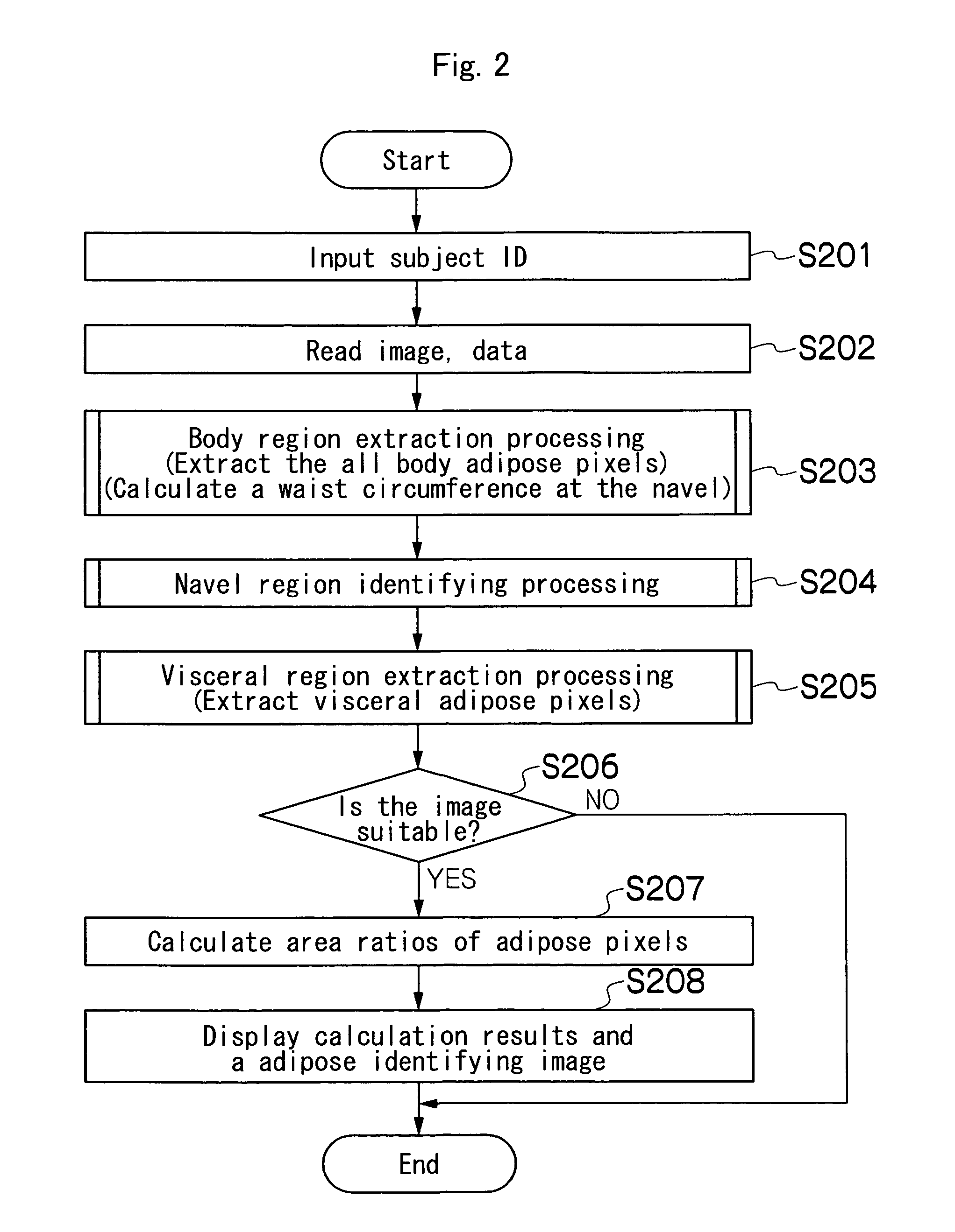
[0057]FIG. 2 is a flowchart of a first embodiment for measuring body adipose of a subject using the medical image diagnosing support apparatus 10 configured as described above. The CPU 12 controls the medical image diagnosing support apparatus 10 according to this flowchart.

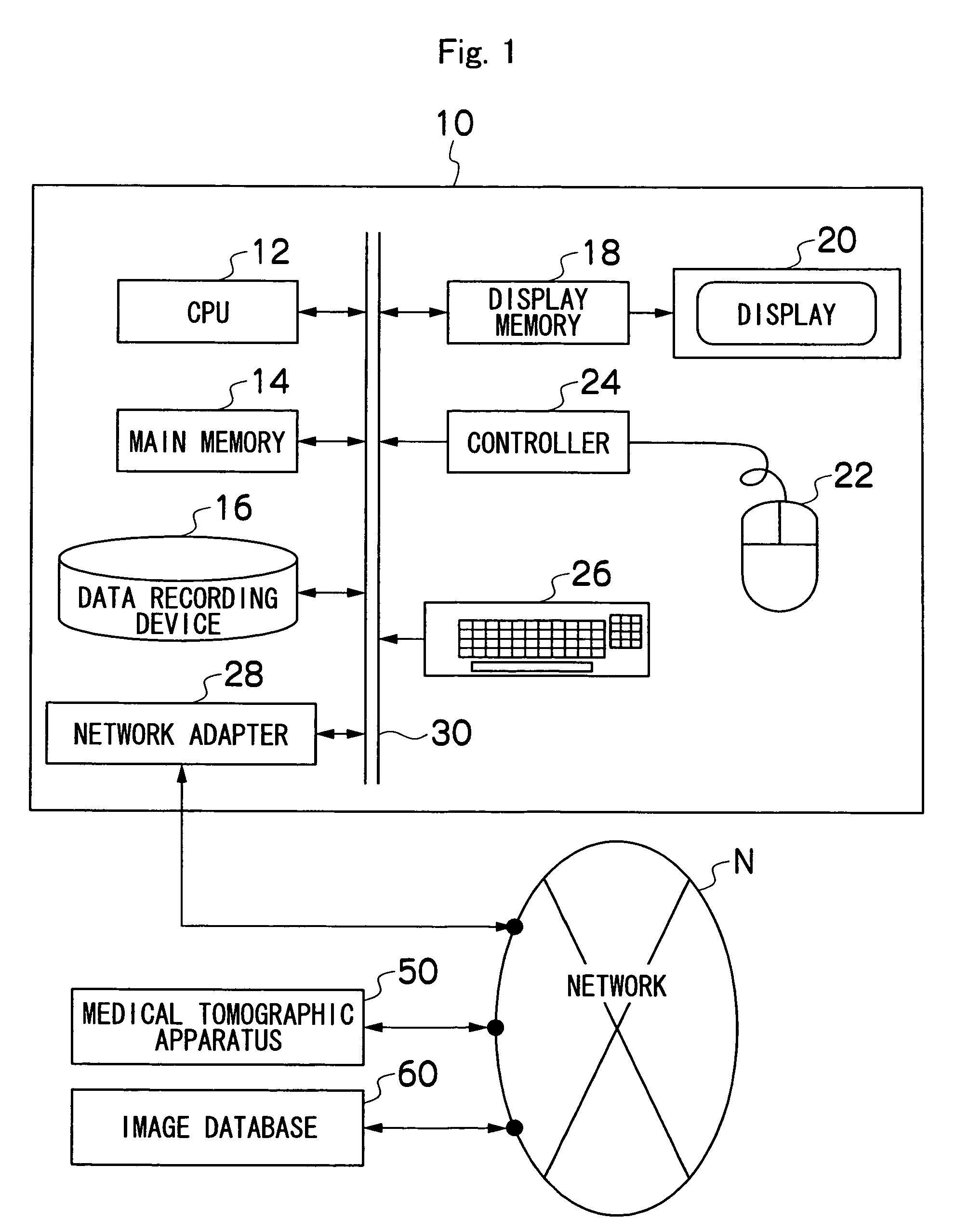
[0058]First, an ID input screen of the subject is displayed on the display 20, and an operator inputs an ID number of the subject of diagnostic processing (S201). Then, based on the input ID number of the subject, image data for body adipose measurement processing is read from the data recording device 16, the medical tomographic apparatus 50, or the image database 60 (S202). Image data acquired by a medical tomographic apparatus such as an X-ray CT apparatus or an MRI apparatus is available, and CT image data acquired by the X-ray CT apparatus will be described by way of an example.

[0059]Next, body region extraction processing (S203), navel region identifying processing (S204), and visceral region extraction pro...

third embodiment

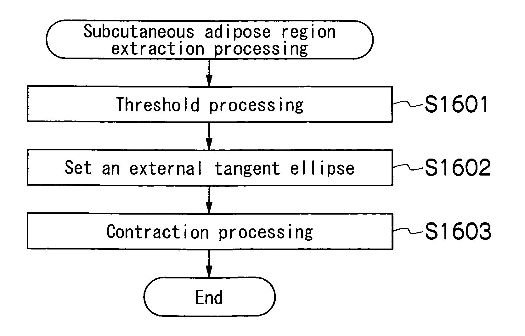
[0071]FIG. 19 is a flowchart of a third embodiment for measuring body adipose of a subject using a medical image diagnosing support apparatus 10, Image input is performed as in S201 and S202 in FIG. 2 (S1901), body region extraction processing is performed like the processing in 5203 in FIG. 2 (S1902), and a navel region is identified like the processing in S204 in FIG. 2 or the processing in 51403 in FIG. 14 (S1903). Then, a preset region is removed from the body region extracted in S1902, and thus an epidermal region, i.e., the region where epidermal tissue exists, is removed from a region for body adipose measurement (S1904). Then, extraction processing of a muscle and bone region is performed like the processing in S701 in FIG. 7 (S1905). Then, navel region removal processing (S 1906) and estimation of the subcutaneous adipose region (S1907) are sequentially performed. Then, adipose region dividing processing is performed like the processing in S205 in FIG. 7 or the processing i...


PUM


Abstract

A medical image diagnosing support apparatus comprises: a first extraction means which extracts a body region of a subject from a tomographic image of the subject acquired by a medical tomographic apparatus; a second extraction means which extracts a non-adipose region from the body region; a third extraction means which extracts a total body adipose region from the body region; a separation means which separates the total body adipose region into a visceral adipose region and a subcutaneous adipose region based on positional information of the non-adipose region; and a display control means which controls of displaying the tomographic image on an image display device with clear indication of the visceral adipose region and the subcutaneous adipose region.

Description

TECHNICAL FIELD[0001]This disclosure relates to a medical image diagnosing support apparatus and method for measuring body adipose of a subject based on a tomographic image of the subject acquired by a medical tomographic apparatus such as an X-ray CT (computed tomography) apparatus or an MRI (magnetic resonance imaging) apparatus, More particularly, the present invention relates to a medical image diagnosing support apparatus and method that can automatically separately measure subcutaneous adipose and visceral adipose using information on a periphery of adipose tissue such as muscle tissue of the subject.BACKGROUND ART[0002]Japanese Patent Application Laid-Open No. 2002-222410 discloses an image diagnosing apparatus that measures body adipose of a subject based on a tomographic image of the subject acquired from a medical tomographic apparatus. The image diagnosing apparatus sets a boundary between subcutaneous adipose and visceral adipose with respect to a peripheral edge of a mu...

Claims


Application Information

Patent Timeline
Patent Type & Authority Patents(United States)
IPC IPC(8): G06K9/00A61B6/03G06T5/00G06T7/00
CPCG06T7/0012G06T7/0081G06T2207/10072G06T2207/20132G06T2207/30004G06T7/11
Inventor NAKASHIMA, KUNIYOSHINAKAGAWA, TORU
Owner FUJIFILM HEALTHCARE CORP
Why Eureka
Try Eureka