
Membrane type-1 matrix metalloprotein inhibitors and uses thereof
a technology of membrane type 1 and metalloprotein, which is applied in the direction of peptide/protein ingredients, peptide sources, drug compositions, etc., can solve the problems of destroying healthy tissue, pain, stiffness, limited motion, etc., and achieves the effect of reducing the frequency or severity
- Summary
- Abstract
- Description
- Claims
- Application Information
AI Technical Summary
Benefits of technology
Problems solved by technology
Method used
Image

Examples
example 1
Generation of MT1-MMP Derived Peptides
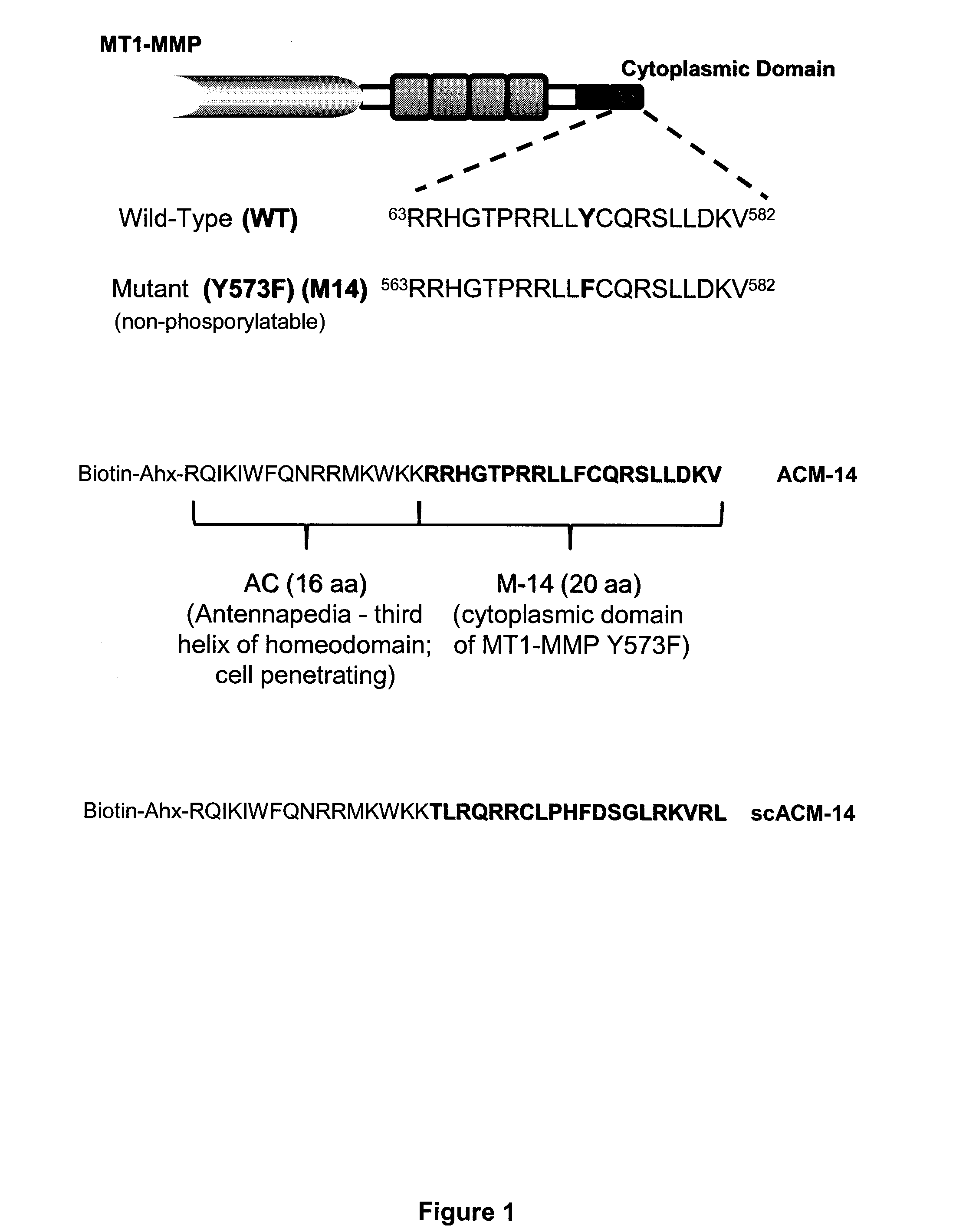
[0126]In performing the experiments described in the examples below, the following peptides were generated. First, a peptide having the sequence of the cytoplasmic domain of MT1-MMP (RRHGTPRRLLYCQRSLLDKV; SEQ ID NO:117), as well as a nonphosphorylatable form of this peptide (RRHGTPRRLLFCQRSLLDKV; SEQ ID NO:118), which has a phenylalanine in place of a tyrosine at the position corresponding to amino acid 573 of the human MT1-MMP sequence were generated. The nonphosphorylatable peptide has been termed “M-14.”
[0127]In addition, peptides having the M-14 sequence fused to the cell penetrating third helix of the homeodomain of Antennapedia protein (RQIKIWFQNRRMKWKK; SEQ ID NO:119) were generated. This fusion peptide is called ACM-14 and has the sequence Biotin-Ahx-RQIKIWFQNRRMKWKK-RRHGTPRRLLFCQRSLLDKV (SEQ ID NO:176). For use as a control, a version of the fusion peptide, where the cytoplamsic MT1-MMP domain sequence is scrambled was generated. This p...
example 2
Expression of Y573F MT1-MMP Inhibits Tumor Growth
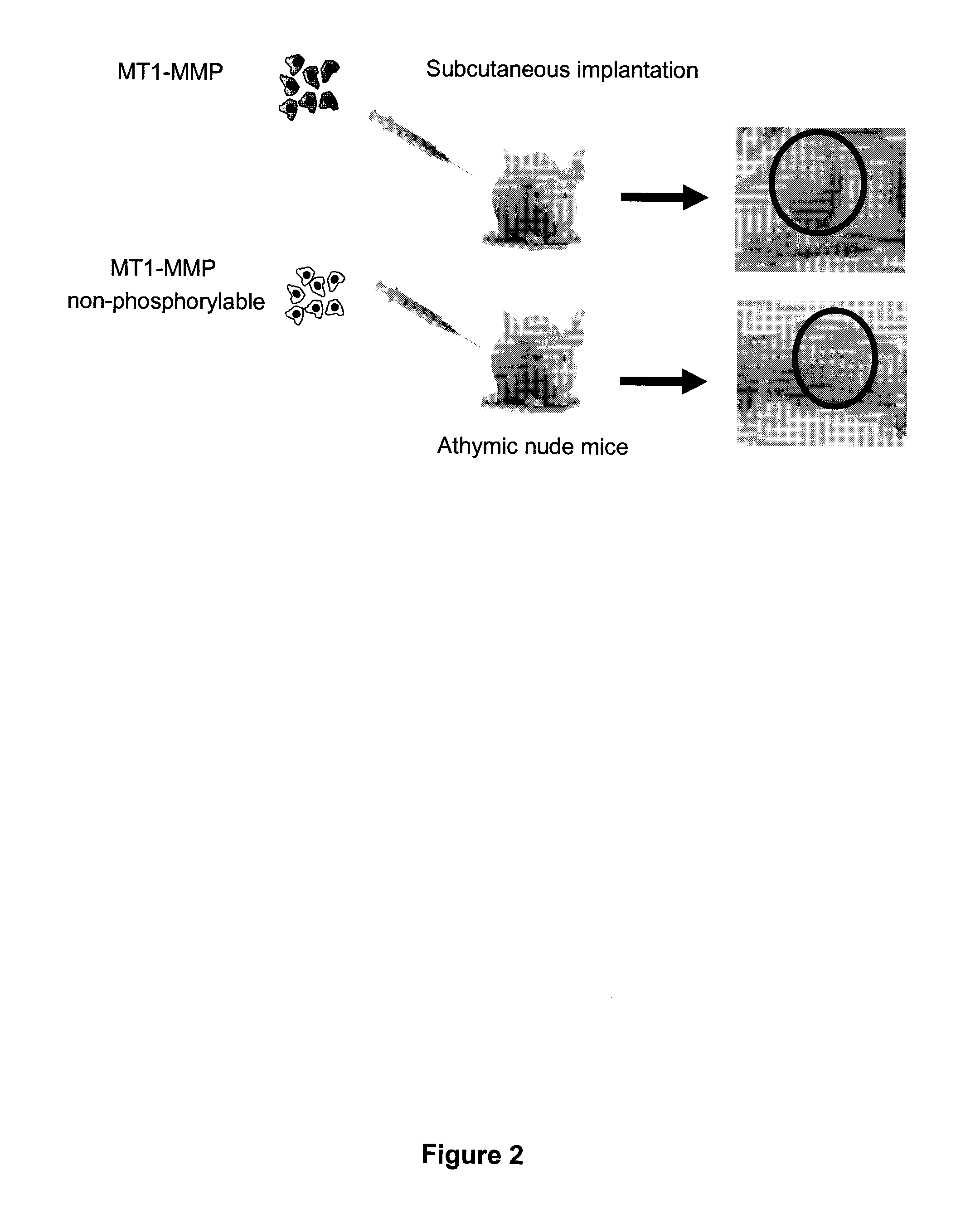
[0128]HT1080 fibrosarcoma cells were stably transfected with either WT MT1-MMP or Y573F MT1-MMP. HT1080 cells are very aggressive cancer cells that express elevated levels of MT1-MMP. Both groups of cells were transplanted subcutaneously into athymic nude mice, and tumor growth was monitored. As shown in FIG. 2, cells expressing the cytoplasmic domain of MT1-MMP exhibited significant tumor growth, whereas no tumor growth was observed in the mice that received cells expressing Y573F mutant.
example 3
ACM-14 and scACM-14 are efficiently Taken Up by Fibrosacrcoma Cells
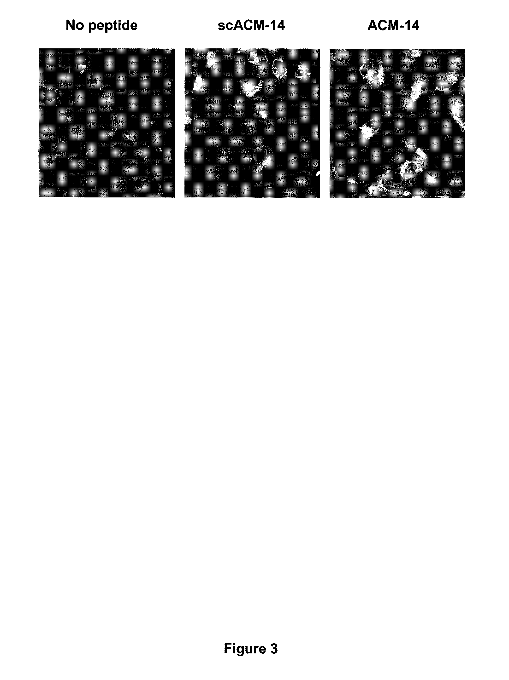
[0129]To determine whether ACM-14 and scACM-14 were capable of entering tumor cells, HT-1080 fibrosarcoma cells were incubated with each peptide (1 μM) for one hour, and peptide uptake was analyzed by immunofluorescence and confocal microscopy. As shown in FIG. 3, both ACM-14 and its scrambled version (scACM-14) were visualized in the cells, indicated efficient and rapid cellular uptake.
PUM
| Property | Measurement | Unit |
|---|---|---|
| pH | aaaaa | aaaaa |
| weight | aaaaa | aaaaa |
| time | aaaaa | aaaaa |
Abstract
Description
Claims
Application Information