

Replication factor C-40 (RFC40/RFC2) as a prognostic marker and target in estrogen positive and negative and triple negative breast cancer
a breast cancer and recombination factor technology, applied in the field of breast cancer, can solve the problems of huge void for patients' therapies, inability to respond to tnbc therapies, and most remarkable and challenging steps, and achieve the effect of modulating cell division and/or proliferation
- Summary
- Abstract
- Description
- Claims
- Application Information
AI Technical Summary
Benefits of technology
Problems solved by technology
Method used
Image

Examples
example 1
RFC40 is a Molecular Marker for Breast Cancer
[0184]Cancers with gene copy number amplification or chromosome polysomy usually are the most aggressive types, often associated and correlated with invasion, metastasis and recurrence, and cannot be corrected at the chromosomal level (Mizutani T et al (1993) Cancer 72(7):2083-2088; Birkeland E et al (2012) Br J Cancer 107(12):1997-2004; Vlajnic T et al (2011) Modern Pathology 24:1404-1412; Lundgren K et al (20120 Breast Cancer Res 14(2):R57). In such cases a strategy for effective cancer treatment is early detection and development of potent chemotherapeutic drugs that will selectively target protein(s) in the cancerous cells.
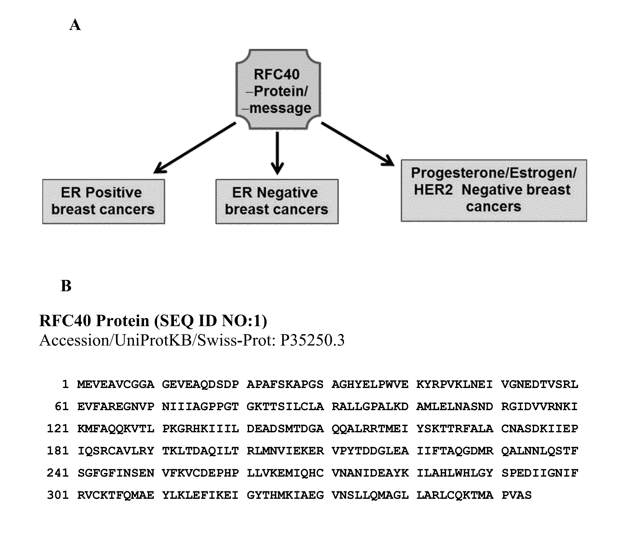
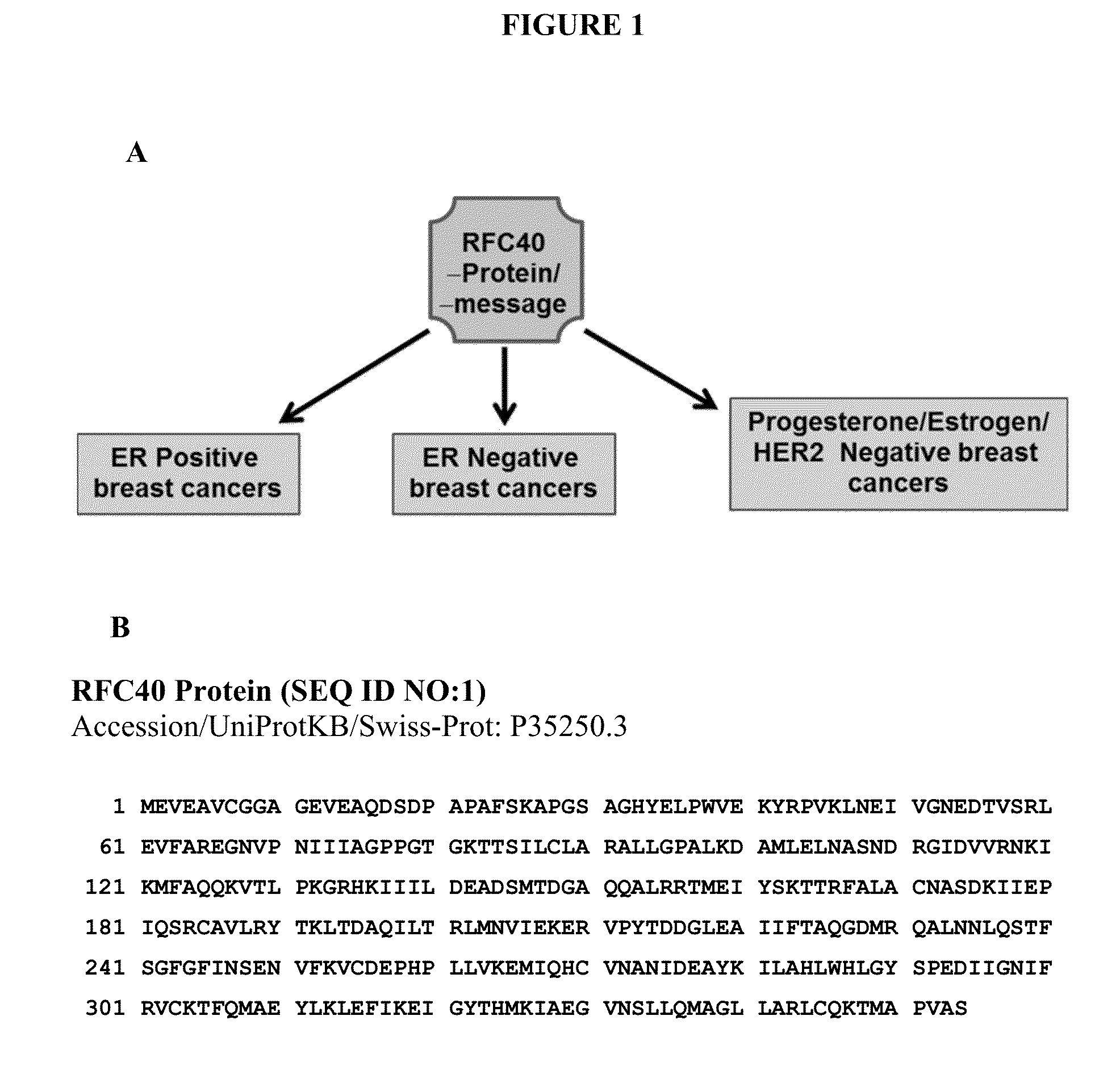
[0185]In this disclosure, we validate RFC40 as a non-receptor based molecular marker for breast cancers specifically, estrogen positive (ER positive), estrogen negative (ER negative), and progesterone, estrogen and human epidermal growth factor receptor 2-HER2 negative or triple negative breast cancers (TNBC). The a...
example 2
RFC40 is Localized in the Nucleus
[0197]It was determined that the pre-dominant localization of RFC40 in the nucleus is an indicator of the highly proliferative state of cancer cells. MCF10A, MCF7 and MDA-MB-231 cells were fixed and subjected to immunofluorescence microscopy using polyclonal anti-RFC40 antibody followed by incubation with Alexa-488-conjugated secondary antibodies for 1 h as described previously (Gupte R et al (2005) Cancer Biology and Therapy 4(4):429-437). Images of the stained sections were collected using a Nikon A1 microscope with Plan ×40 / NA 0.25 Phi objective. Nuclear staining of RFC40 was more pronounced and significant in the breast cancer cells versus normal-cancerous breast cells (FIG. 7A). MCF10A, MCF7 and MDA-MB-231 cells were then subjected to flow cytometric analyses using DAPI (MCF10A) and PI (MCF7 and MDA-MB-231), respectively. Histograms representing the percent of cells in G1, S and G2 phases respectively demonstrate significantly higher percentage ...
example 3
The RFC40 Gene is Amplified in Breast Cancer
[0199]Over-expression of any protein(s) in cancer can occur due to aberrant amplification in its gene copy numbers in addition to up-regulation of its message and hence protein. Interestingly, amplification of RFC40 gene copy numbers as been previous demonstrated in glioblastomas (Nakahara Y et al (2004) Neuro Oncol 6(4):281-9; Suzuki T et al (2004) Brain Tumor Pathol 21(1):27-34). However, whether RFC40 gene copy numbers are increased in breast cancers has not been investigated. We propose that RFC40 gene copy number is amplified in breast cancers, as illustrated in the schematic (FIG. 9).
[0200]We performed qualitative gene copy number analyses for the RFC40 promoter region located on the chromosome 7 at q11.23 position. We isolated genomic DNA from Adult normal breast tissue, MCF10A and MDA-MB-468 cells and performed qualitative PCR to amplify a 206 bp fragment on the RFC40 promoter and analyzed it on 4% agarose gel. The data suggested t...
PUM
 Abstract
Description
Claims
Application Information