Systems for temperature-controlled ablation using radiometric feedback

a radiometric feedback and temperature control technology, applied in the field of systems and methods for measuring and controlling temperature during tissue ablation, can solve the problems of obscuring any useful information about clinicians having no useful feedback regarding the temperature of the tissue, etc., to achieve the effect of sufficient ablation

Active Publication Date: 2016-01-05
CORAL SAND BEACH LLC +1
View PDF369 Cites 173 Cited by
  • Summary
  • Abstract
  • Description
  • Claims
  • Application Information

AI Technical Summary

Benefits of technology

[0014]In view of the foregoing, it would be desirable to provide apparatus and methods for treating living tissue that employs a radiometer for temperature measurement, and a temperature control subsystem that uses feedback from the radiometer to regulate the power of ablation energy being applied to the tissue. In accordance with one aspect of the invention, systems and methods are provided for radiometrically measuring temperature during RF ablation, i.e., calculating temperature based on signal(s) from a radiometer. Unlike standard thermocouple techniques used in existing commercial ablation systems, a radiometer may provide useful information about tissue temperature at depth—where the tissue ablation occurs—and thus provide feedback to the clinician about the extent of tissue damage as the clinician ablates a selected region of the heart muscle. Furthermore, the temperature control subsystem may automatically regulate the power of the ablation energy applied to the tissue based on the tissue temperature, so as to maintain the tissue at the desired temperature and for the desired amount of time to achieve sufficient ablation.
[0017]The computer-readable medium preferably stores instructions that cause the processor to execute the step of calculating a temperature adjacent to the ICT based on the digital radiometer signal, the digital thermocouple signal, and the operation parameters. This temperature is expected to provide significantly more accurate information about lesion quality and temperature at depth in the tissue than would a temperature based solely on a thermocouple readout. The computer-readable medium may further store instructions for causing the processor to cause the temperature display to display the calculated temperature, for example so that the clinician may control the time period for ablation responsive to the displayed temperature. The computer-readable medium may further store instructions for causing the processor to close the patient relay, such that the patient relay passes ablative energy received on the second I / O port, from the electrosurgical generator, to the first I / O port, to the ICT. Note that the instructions may cause the processor to maintain the patient relay in a normally closed state, and to open the patient relay upon detection of unsafe conditions.

Problems solved by technology

Although commercially available ablative tips may include thermocouples for providing temperature feedback via a digital display, such thermocouples typically do not provide meaningful temperature feedback during irrigated ablation.
Moreover, for procedures in which the surface of the tissue is cooled with an irrigant, the thermocouple will measure the temperature of the irrigant, thus further obscuring any useful information about the temperature of the tissue, particularly at depth.
As such, the clinician has no useful feedback regarding the temperature of the tissue as it is being ablated or whether the time period of the ablation is sufficient.
Because the clinician lacks such information, the clinician furthermore cannot regulate the power of the ablation energy so as to heat or cool the tissue to the desired temperature for a sufficient period of time.
Accordingly, it may only be revealed after the procedure is completed—for example, if the patient continues to experience cardiac arrhythmias—that the targeted aberrant pathway was not adequately interrupted.
In such a circumstance, the clinician may not know whether the procedure failed because the incorrect region of tissue was ablated, because the ablative tip was not actuated for a sufficient period of time to destroy the aberrant pathway, because the ablative tip was not touching or sufficiently touching the tissue, because the power of the ablative energy was insufficient, or some combination of the above.
Upon repeating the ablation procedure so as to again attempt to treat the arrhythmia, the clinician may have as little feedback as during the first procedure, and thus potentially may again fail to destroy the aberrant pathway.
Additionally, there may be some risk that the clinician would re-treat a previously ablated region of the endocardium and not only ablate the conduction pathway, but damage adjacent tissues.
However, there is again insufficient feedback to assist the clinician in determining whether any of those ablated regions are sufficiently destroyed.
Despite the promise of precise temperature measurement sensitivity and control offered by the use of radiometry, there have been few successful commercial medical applications of this technology.
One drawback of previously-known systems has been an inability to obtain highly reproducible results due to slight variations in the construction of the microwave antenna used in the radiometer, which can lead to significant differences in measured temperature from one catheter to another.
Problems also have arisen with respect to orienting the radiometer antenna on the catheter to adequately capture the radiant energy emitted by the tissue, and with respect to shielding high frequency microwave components in the surgical environment so as to prevent interference between the radiometer components and other devices in the surgical field.
Acceptance of microwave-based hyperthermia treatments and temperature measurement techniques also has been impeded by the capital costs associated with implementing radiometric temperature control schemes.
Radiofrequency ablation techniques have developed a substantial following in the medical community, even though such systems can have severe limitations, such as the inability to accurately measure tissue temperature at depth, e.g., where irrigation is employed.
However, the widespread acceptance of RF ablation systems, extensive knowledge base of the medical community with such systems, and the significant cost required to changeover to, and train for, newer technologies has dramatically retarded the widespread adoption of radiometry.

Method used


Image

  • Systems for temperature-controlled ablation using radiometric feedback
  • Systems for temperature-controlled ablation using radiometric feedback
  • Systems for temperature-controlled ablation using radiometric feedback

Examples


Embodiment Construction

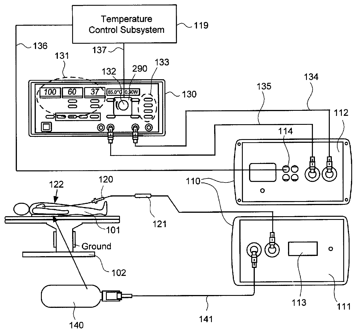
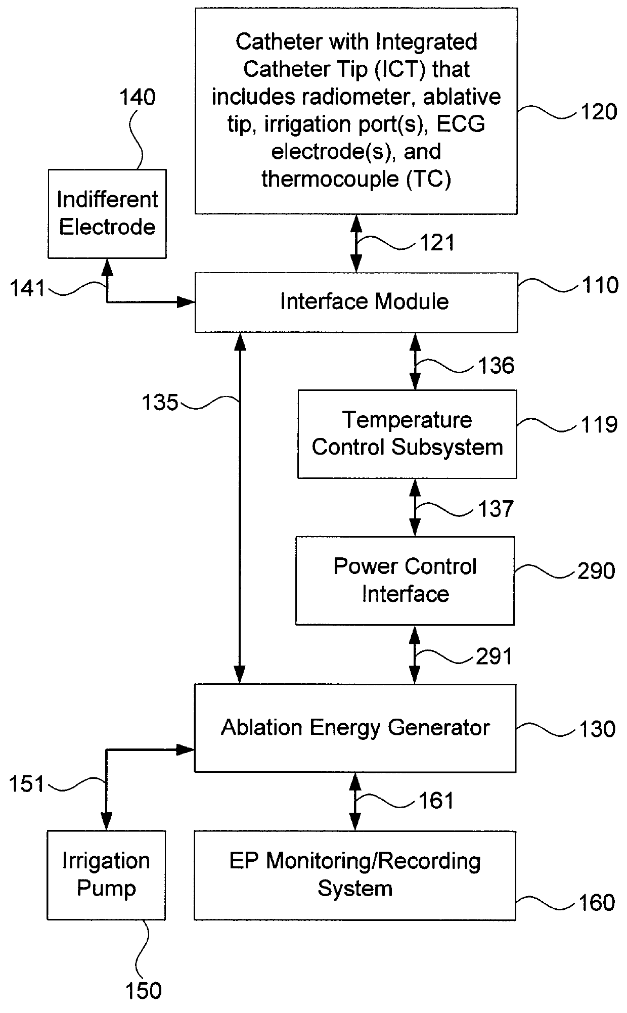
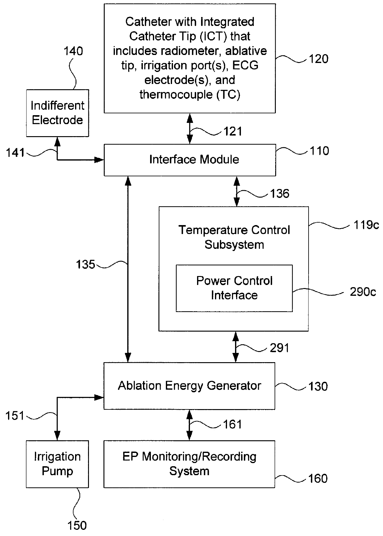
[0035]Embodiments of the present invention provide systems and methods for radiometrically measuring temperature during ablation, in particular cardiac ablation, and for automatically regulating the power of ablation energy based on same. As noted above, commercially available systems for cardiac ablation may include thermocouples for measuring temperature, but such thermocouples may not adequately provide the clinician with information about tissue temperature. Thus, the clinician may need to make an “educated guess” about whether a given region of tissue has been sufficiently ablated to achieve the desired effect. By comparison, calculating a temperature based on signal(s) from a radiometer is expected to provide accurate information to the clinician about the temperature of tissue at depth, even during an irrigated procedure. Furthermore, a temperature control subsystem may be employed that monitors the calculated temperature, and automatically regulates or controls the power of ...


PUM


Abstract

The present invention provides systems and methods for temperature-controlled ablation using radiometric feedback. An interface module may include (a) a processor; (b) a first input / output (I / O) port to receive digital radiometer and digital thermocouple signals from an integrated catheter tip (ICT); (c) a second I / O port to receive ablative energy from a generator; (d) a temperature display; (e) a patient relay; (f) a computer-readable medium storing instructions for causing the processor to: (i) calculate a temperature adjacent to the ICT based on the digital radiometer and thermocouple signals and operation parameters; (ii) cause the temperature display to display the calculated temperature; and (iii) close the patient relay to pass ablative energy received on the second I / O port to the first I / O port; and (g) a temperature control subsystem to regulate the ablative power based on the calculated temperature.

Description

FIELD OF THE INVENTION[0001]This application generally relates to systems and methods for measuring and controlling temperature during tissue ablation.BACKGROUND OF THE INVENTION[0002]Tissue ablation may be used to treat a variety of clinical disorders. For example, tissue ablation may be used to treat cardiac arrhythmias by destroying aberrant pathways that would otherwise conduct abnormal electrical signals to the heart muscle. Several ablation techniques have been developed, including cryoablation, microwave ablation, radio frequency (RF) ablation, and high frequency ultrasound ablation. For cardiac applications, such techniques are typically performed by a clinician who introduces a catheter having an ablative tip to the endocardium via the venous vasculature, positions the ablative tip adjacent to what the clinician believes to be an appropriate region of the endocardium based on tactile feedback, mapping electrocardiogram (ECG) signals, anatomy, and / or fluoroscopic imaging, ac...

Claims


Application Information

Patent Timeline
Patent Type & Authority Patents(United States)
IPC IPC(8): A61B18/14A61B18/08A61B18/12A61B18/18A61B17/00A61N7/02A61B18/00A61B18/02
CPCA61B18/082A61B18/12A61B18/02A61B18/1206A61B18/1492A61B18/1815A61B2017/0007A61B2017/00486A61B2018/00357A61B2018/00577A61B2018/00642A61B2018/00678A61B2018/00708A61B2018/00714A61B2018/00797A61B2018/00821A61B2018/00839A61B2218/002A61N7/022A61N7/02
Inventor MCCARTHY, JOHN F.LENIHAN, TIMOTHY J.KANOWSKY, ERIC R.ALLISON, ROBERT C.
Owner CORAL SAND BEACH LLC
Why Eureka
Try Eureka