Method for tissue resection

a tissue resection and tissue technology, applied in the field of tissue resection, can solve the problems of small stress on the patient, limitation of the size of the resectable lesioned part, and large stress on the patien

Active Publication Date: 2017-07-18
OLYMPUS CORP
View PDF6 Cites 23 Cited by
  • Summary
  • Abstract
  • Description
  • Claims
  • Application Information

AI Technical Summary

Problems solved by technology

In the laparotomy method, a wide range of resection can also be easily performed, but the stress to a patient is large.
Meanwhile, in the method using an endoscope, the stress to a patient is small, but there is a limitation to the size of a resectable lesioned part.
However, it is a natural phenomenon that an operation having a high degree of difficulty, such as locking a stomach wall to an abdominal wall and lifting the stomach, is required.

Method used


Image

  • Method for tissue resection
  • Method for tissue resection
  • Method for tissue resection

Examples


first embodiment

Method

[0043]A first embodiment of the present invention will be described with reference to FIGS. 1 to 7. In the present embodiment, a tissue resection method related to the present invention will be described taking as an example a case where a given region of tissue including a lesioned part is resected over all layers, using the large intestine serving as a hollow organ as a target.

[0044]In the following description, a surgeon that approaches resection target tissue from an inner cavity side of the large intestine is called a first surgeon, and a surgeon that approaches the resection target tissue from an abdominal cavity (body cavity) side is called a second surgeon.

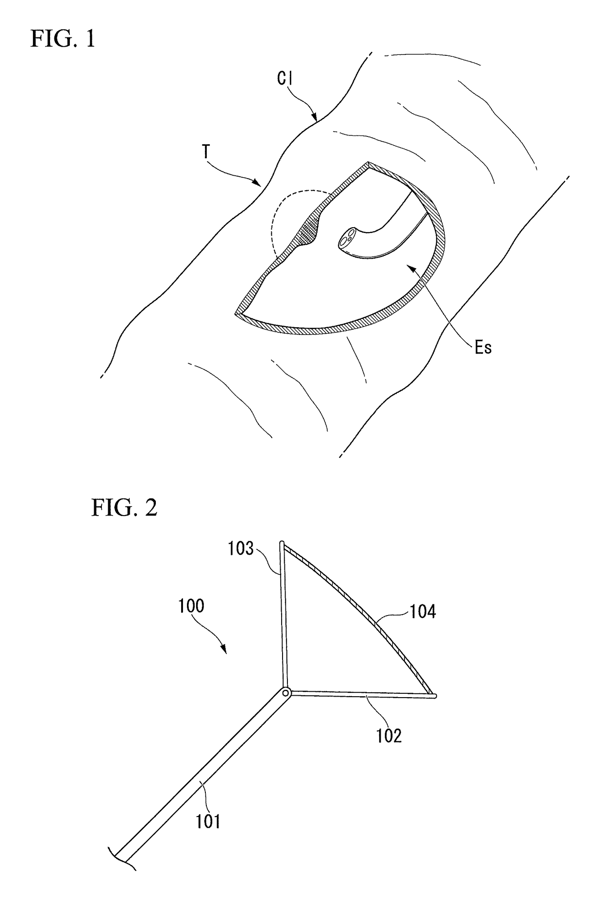
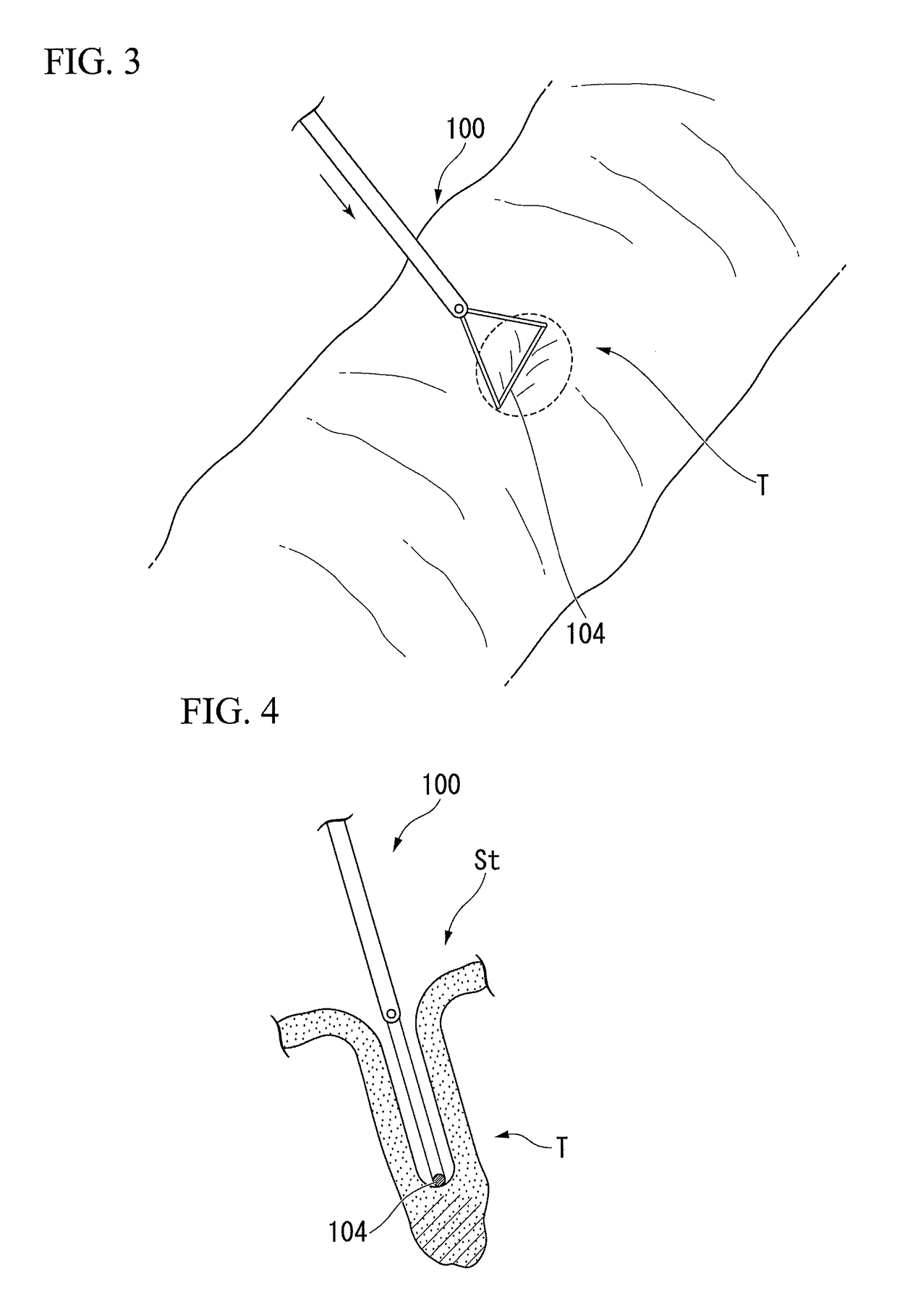
[0045]First, as shown in FIG. 1, the first surgeon introduces an observation portion, such as an endoscope Es, into the large intestine Cl, and observes the inside of the large intestine Cl with the observation portion, and specifies the position and the range of resection target tissue T (first step).

[0046]After the...

second embodiment

System

[0063]Next, a second embodiment of the present invention will be described with reference to FIGS. 8 to 23. In the present embodiment, a tissue resection system that can suitably perform the tissue resection method of the present invention will be described.

[0064]In the following description, the same components as those already described will be designated by the same reference numerals, and duplicate description thereof will be omitted.

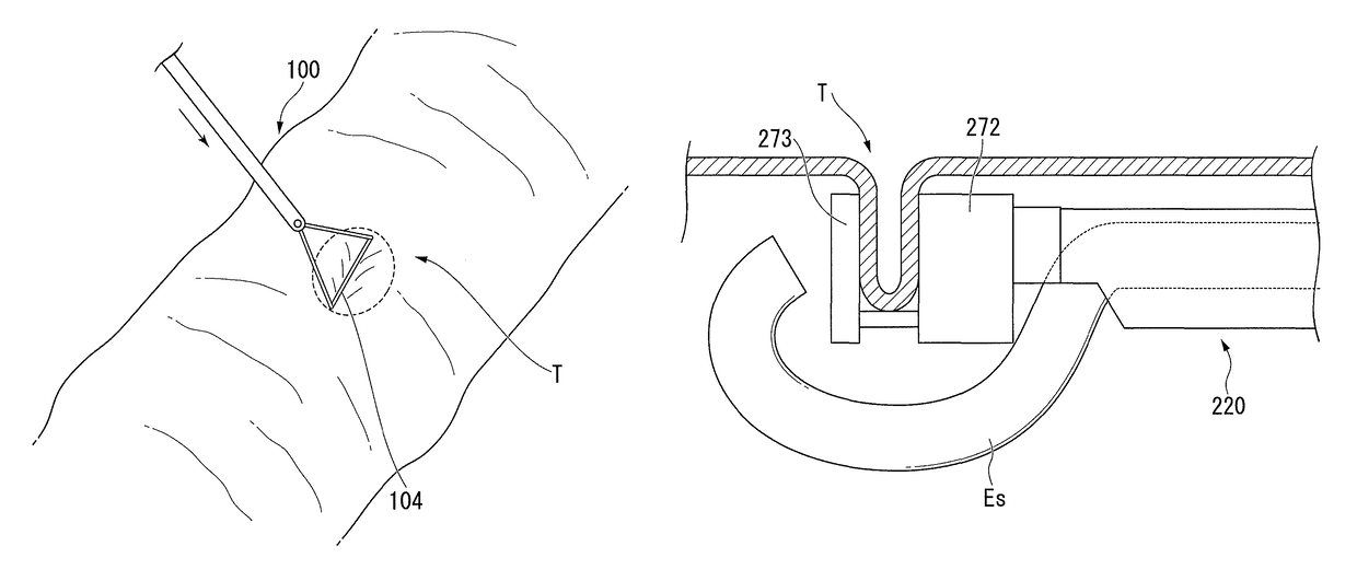
[0065]FIG. 8 is a view shown the tissue resection system 200) of the present embodiment. The tissue resection system 200 includes a tissue pushing tool and a resection anastomosis device 210. The above-described tissue pushing tool 100 is shown as an example of the tissue pushing tool.

[0066]The resection anastomosis device 210 of the present embodiment will be described. The resection anastomosis device 210 includes a tubular insertion section 220 through which an endoscope is inserted, a treatment section 230 that is provided at a distal end ...

modified example 1

[0085]FIG. 16 is a view shown the treatment section in a resection anastomosis device 250 of Modified example 1 in the same aspect as FIG. 10. In the resection anastomosis device 250, only one advance / retraction shaft 252 connected to the anvil portion (not shown) is provided. The sectional shape of the advance / retraction shafts 252, as shown in FIG. 16, is formed in a substantial U shape that opens toward the central axis X1 of the main body 231, and the tissue pressing line 13 is specified by both ends 252a (tissue pressing portion) and 252b (tissue pressing portion) that are separated from each other in U shape.

[0086]The same effects as those of the above-described resection anastomosis device 210 are also exhibited in such a configuration.

[0087]Moreover, the rigidity of the advance / retraction shaft 252 can be enhanced. Additionally, since the number of advance / retraction shafts to be operated is one, an operating mechanism for the advance / retraction shaft can be simply configure...


PUM


Abstract

A tissue resection method of resecting a portion of a hollow organ includes a first step of specifying a position and a range of a resection target tissue from the inside of the hollow organ and showing the position and the range in a visually recognizable manner from the inside of a body cavity; a second step of bringing a bioabsorbable member introduced into the body cavity into contact with the resection target tissue and pressing the resection target tissue, thereby inverting the resection target tissue to the inside of the hollow organ; and a third step of resecting the inverted resection target tissue over all layers from the inside of the hollow organ and performing anastomosis of a hole formed in the hollow organ from the inside of the hollow organ.

Description

TECHNICAL FIELD[0001]The present invention relates to a method for tissue resection.BACKGROUND ART[0002]As methods of resecting a portion of a hollow organ, such as an alimentary canal, a method of performing a laparotomy that largely incises the abdomen, and a method of performing resection without incising the abdomen using an endoscope or the like are known.[0003]In the laparotomy method, a wide range of resection can also be easily performed, but the stress to a patient is large. Meanwhile, in the method using an endoscope, the stress to a patient is small, but there is a limitation to the size of a resectable lesioned part.[0004]As described above, both the laparotomy method and the method using an endoscope or the like have disadvantages. Therefore, tissue resection methods that can perform resection in a range wider than the method using an endoscope or the like and that are less invasive to a patient than the laparotomy method are required.[0005]In relation to this, operativ...

Claims


Application Information

Patent Timeline
Patent Type & Authority Patents(United States)
IPC IPC(8): A61B17/11A61B17/115A61B17/00A61B17/32A61B17/072A61B90/00
CPCA61B17/1114A61B17/00234A61B17/07207A61B17/115A61B17/320016A61B90/361A61B90/37A61B2017/00004A61B2017/00818A61B2017/07221A61B2090/0807A61B2090/0817
Inventor MOTAI, KOSUKE
Owner OLYMPUS CORP
Why Eureka
Try Eureka