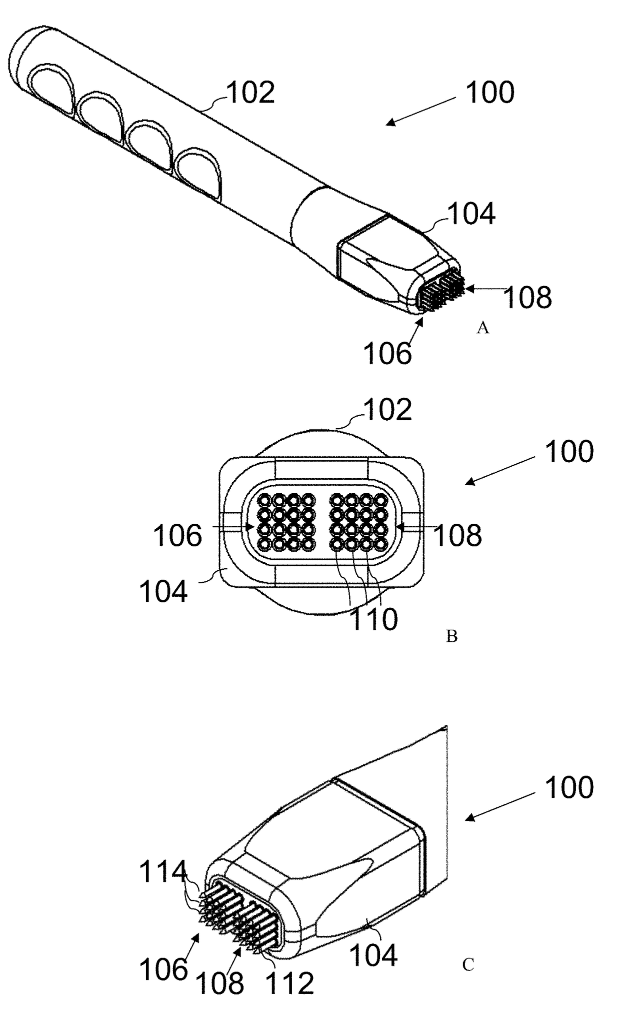
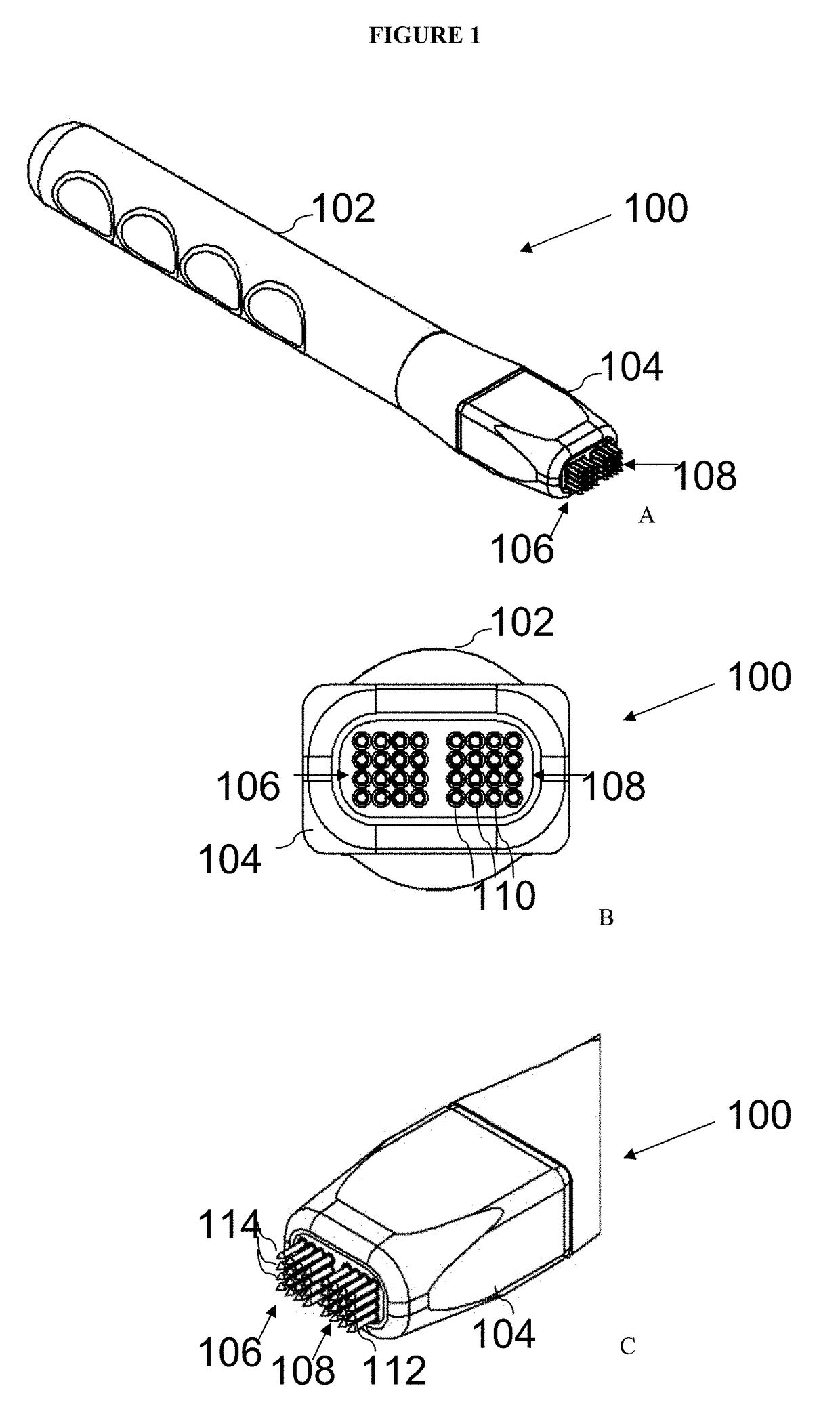
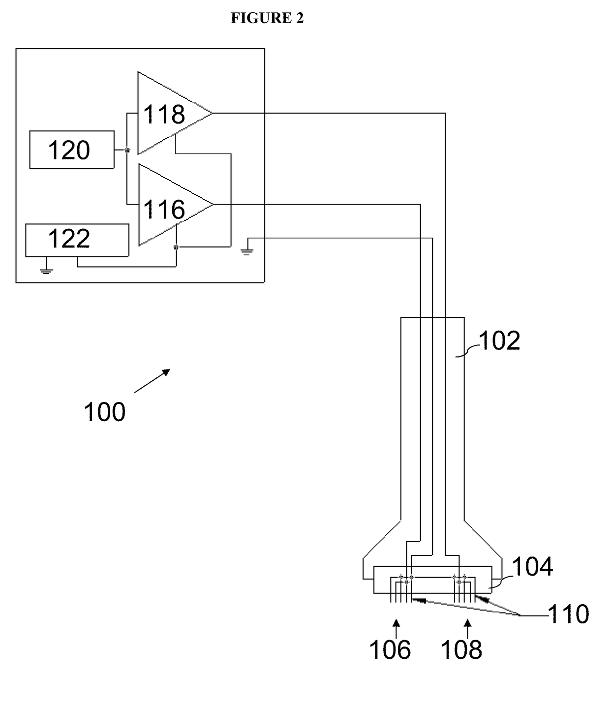
Miniminally invasive dermal electroporation device
a dermal electroporation device, minimally invasive technology, applied in the direction of antigenic vaccination, dna/rna vaccination, therapy, etc., can solve the problems of ineffective delivery of naked need of dna vaccine delivery, and inability to deliver dna through intramuscular injection
- Summary
- Abstract
- Description
- Claims
- Application Information
AI Technical Summary
Problems solved by technology
Method used
Image

Examples
example 1
Effect of Electrode Composition on Transfection Efficiency
[0078]To address the effect of electrode material on reporter gene localization, transfection efficiency was compared for two minimally invasive devices (MID) with different electrode compositions (gold and stainless steel). This comparison was to assess whether a cheaper alternative (stainless steel) to gold electrodes could be used while still maintaining transfection efficacy. The electrode composition was easily tested, because the gold-plated electrodes were easily removed from their sockets and replaced with stainless steel electrodes of the same gauge and length. This produced an identical electrode head differing only in the electrode composition.
[0079]The experimental outline is detailed in Table 1.
[0080]
TABLE 1DNANumber ofBiopsyNumber ofElectrodeDNAConcentrationTreatmentRemoval TimeAnimalsCompositionDelivered(mg / ml)Sites(hours)RequiredFinal AnalysisGold-pgWIZ-0.51012, 24, 484GrossplatedGFP11012, 24, 48Visualization / ...
example 2
Effect of Electrode Spacing on Transfection Efficiency
[0084]To assess the effect of electrode spacing on transfection efficiency and reporter gene localization, a 1 mm spaced circuit board was created and fitted in a head-piece housing and compared with a similar MID with a 1.5 mm spaced circuit board. To ensure that the surface treatment area remained the same between the two hand pieces at the different spacings, an additional row of electrodes was added to the 1 mm spaced circuit board. Therefore, a 1.5 mm spacing hand piece with 4×4 rows of electrodes (16 electrodes) was compared to a 1 mm spacing hand piece with 5×5 rows of electrodes (25 electrodes). As such, each hand piece had an approximate treatment surface area of approximately 4-4.5 mm2. FIG. 6A shows a photograph of both hand pieces from a side perspective. The top hand piece is the 1 mm spacing, and the bottom hand piece is the 1.5 mm spacing. FIG. 6B shows a close-up view of the face of the 1 mm hand piece.
[0085]A ser...
example 3
Effect of Electrode Spacing on Current
[0090]A device as described herein can have the capacity to capture and store all electrical parameters real time as they occur during each electroporation pulse. A series of in vivo expression localization studies were completed, as described in Example 1, to examine current and voltage for electroporation with devices of different electrode spacing and composition. While the applied voltage remained constant (15 volts), the impedance (resistance) and current delivered for each treatment was examined for each electrode spacing and each electrode composition.
[0091]FIG. 8 shows both the resulting impedance (resistance in Ohms) and Current (in milli Amps).
[0092]The current was approximately three times greater in the 1 mm hand piece (average 85 mA) compared to the 1.5 mm hand piece (average 23 mA), yet the applied voltage was the same across all conditions. The increased current in the 1.5 mm hand piece resulted in a large reduction (approximately...
PUM
| Property | Measurement | Unit |
|---|---|---|
| length | aaaaa | aaaaa |
| length | aaaaa | aaaaa |
| distance | aaaaa | aaaaa |
Abstract
Description
Claims
Application Information
 - R&D
- Intellectual Property
- Life Sciences
- Materials
- Tech Scout
- Unparalleled Data Quality
- Higher Quality Content
- 60% Fewer Hallucinations
Browse by: Latest US Patents, China's latest patents, Technical Efficacy Thesaurus, Application Domain, Technology Topic, Popular Technical Reports.
© 2025 PatSnap. All rights reserved.Legal|Privacy policy|Modern Slavery Act Transparency Statement|Sitemap|About US| Contact US: help@patsnap.com