Endoscope system, processor device of endoscope system, and image processing method

a technology of endoscope and processor device, which is applied in the field of endoscope system, endoscope system processor device, image processing method, etc., can solve the problem of not being able to reliably extract a blood vessel to be observed

Active Publication Date: 2018-04-17
FUJIFILM CORP
View PDF8 Cites 162 Cited by
  • Summary
  • Abstract
  • Description
  • Claims
  • Application Information

AI Technical Summary

Benefits of technology

[0019]According to the present invention, since blood vessel extraction processing, which differs depending on the light amount ratio of the first illumination light and the second illumination light to illuminate a subject, is performed, it is possible to reliably extract a plurality of types of blood vessels at different depths even if the light amount ratio is changed.

Problems solved by technology

For this reason, in the blood vessel discrimination method based on the hue disclosed in JP2011-135983A it is not possible to reliably extract a blood vessel to be observed.

Method used


Image

  • Endoscope system, processor device of endoscope system, and image processing method
  • Endoscope system, processor device of endoscope system, and image processing method
  • Endoscope system, processor device of endoscope system, and image processing method

Examples


Embodiment Construction

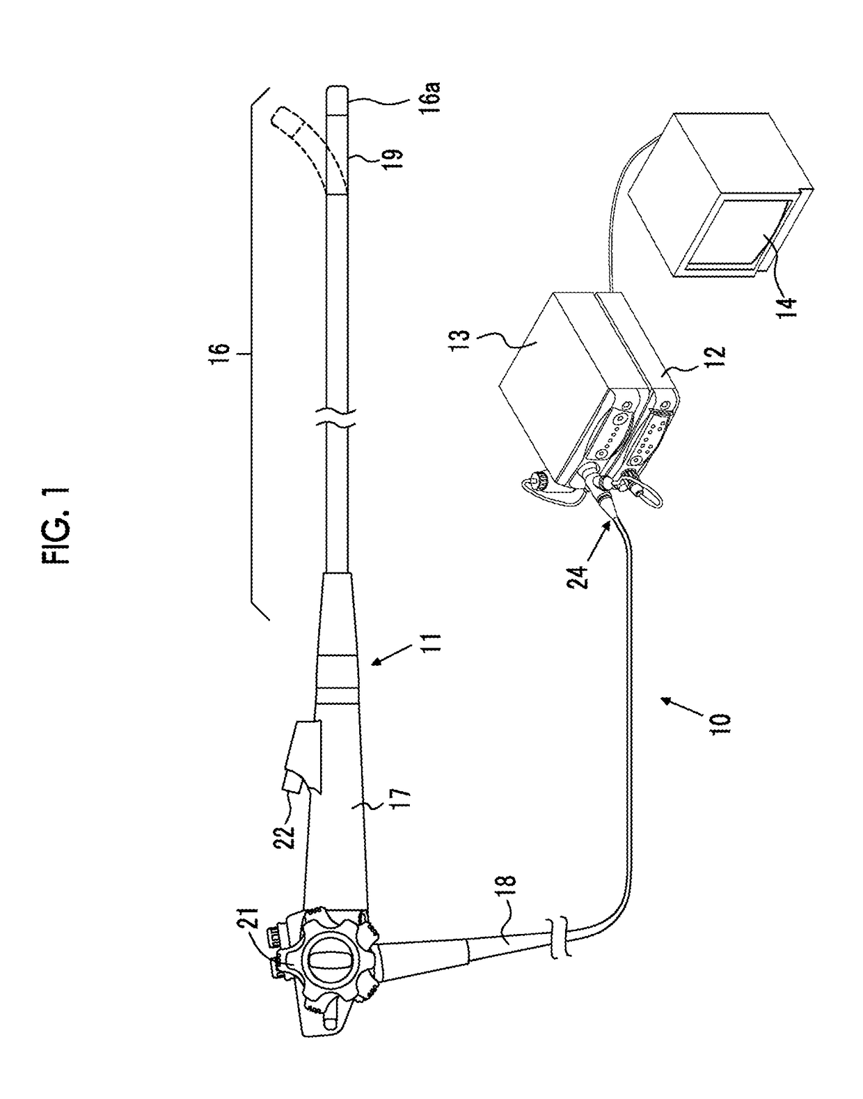
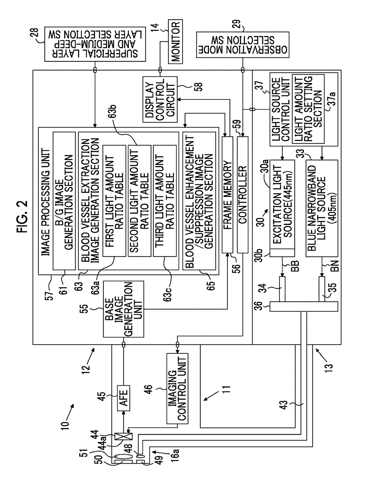
[0042]As shown in FIG. 1, an electronic endoscope system 10 includes an electronic endoscope 11 that images the inside of a subject, a processor device 12 that generates an endoscope image based on a signal obtained by imaging, a light source device 13 that generates light for illuminating the subject, and a monitor 14 that displays an endoscope image. The electronic endoscope 11 includes a flexible insertion unit 16 that is inserted into the body cavity, an operating unit 17 provided at the proximal end of the insertion unit 16, and a universal code 18 that makes a connection between the operating unit 17 and the processor device 12 and the light source device 13.

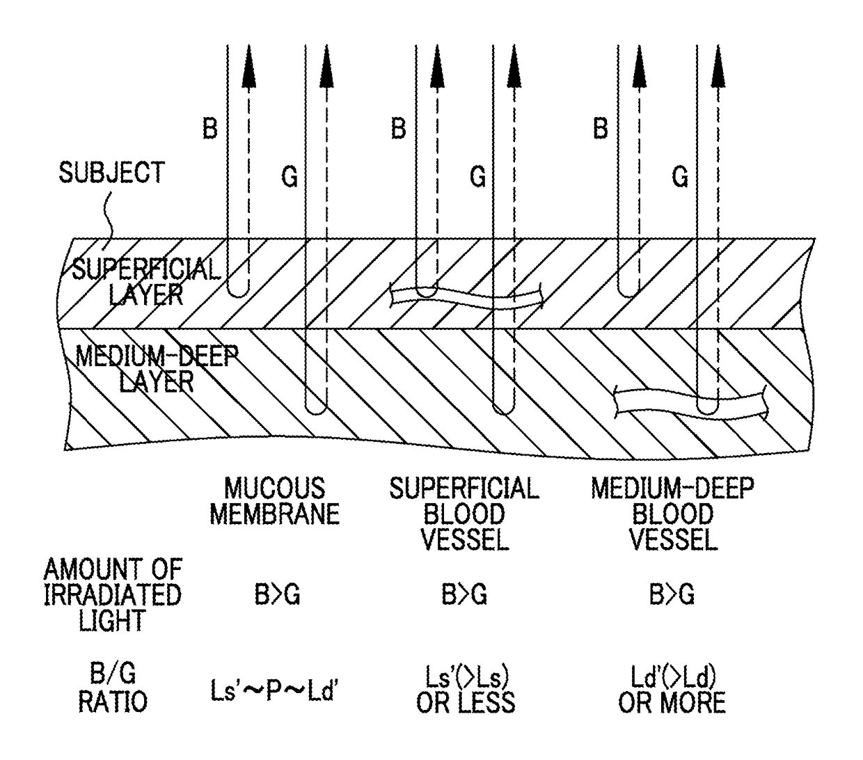
[0043]The electronic endoscope system 10 has a function of generating a superficial blood vessel enhancement image or suppression image, in which a superficial blood vessel of a subject is enhanced / suppressed, and a medium-deep blood vessel enhancement image or suppression image, in which a medium-deep superficial blood ve...


PUM


Abstract

Even if the ratio between blue and green components of illumination light is changed, a plurality of types of blood vessels at different depths are reliably distinguished. A blue signal B, a green signal G, a red signal R is obtained by imaging the subject using a color CCD 44. A B / G image having a B / G ratio is generated. A superficial blood vessel extraction image is obtained by extracting a pixel, in which the B / G ratio is equal to or less than a boundary value Ls between the mucous membrane and the superficial blood vessel, from the B / G image. A medium-deep blood vessel extraction image is obtained by extracting a pixel, in which the B / G ratio is equal to or greater than a boundary value Ld between the mucous membrane and the medium-deep blood vessel. The boundary values Ls and Ld differ depending on the light amount ratio.

Description

CROSS-REFERENCE TO RELATED APPLICATIONS[0001]This application is a Continuation of PCT International Application No. PCT / JP2013 / 050361 filed on Jan. 11, 2013, which claims priority under 35 U.S.C. § 119(a) to Japanese Patent Application No. 2012-013317 filed Jan. 25, 2012. Each of the above applications is hereby expressly incorporated by reference, in its entirety, into the present application.BACKGROUND OF THE INVENTION[0002]1. Field of the Invention[0003]The present invention relates to an endoscope system capable of extracting blood vessels, such as superficial blood vessels and medium-deep blood vessels, from a subject, a processor device of the endoscope system, and an image processing method.[0004]2. Description of the Related Art[0005]In recent medical treatment, diagnosis or the like using an endoscope apparatus has been widely performed. As observation of the inside of a subject using an endoscope apparatus, not only normal observation to observe the subject's inside in it...

Claims


Application Information

Patent Timeline
Patent Type & Authority Patents(United States)
IPC IPC(8): A61B1/04A61B5/00G06T7/00A61B5/02G06K9/00G06T11/00G06T7/90A61B1/00A61B1/06
CPCA61B5/0084A61B1/043A61B1/0638A61B1/0646A61B1/0653A61B5/02007G06K9/0014G06T7/0012G06T7/90G06T11/001A61B1/00009G06T2207/30101A61B1/0005A61B1/063G06T2207/10024G06T2207/10068G06T2207/10152A61B1/000094G06V20/695
Inventor KAKU, TOSHIHIKOIIDA, TAKAYUKI
Owner FUJIFILM CORP
Why Eureka
Try Eureka