

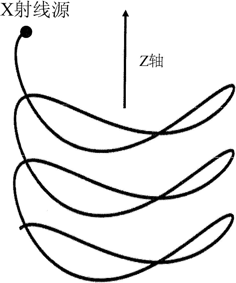
Conical bundle CT reestablishment method based on helix saddle line
A cone-beam and helix technology, applied in the field of biomedical imaging, can solve the problems of one-way movement of X-ray source, fast data acquisition speed, backflow, etc., and achieve the effect of flexible scanning mode and low false detection rate
- Summary
- Abstract
- Description
- Claims
- Application Information
AI Technical Summary
Problems solved by technology
Method used
Image

Examples
Embodiment Construction
[0025] The embodiments of the present invention are described in detail below in conjunction with the accompanying drawings: this embodiment is implemented on the premise of the technical solution of the present invention, and detailed implementation methods and specific operating procedures are provided, but the protection scope of the present invention is not limited to the following the described embodiment.
[0026] Take human angiographic real-time detection as an example. The realization process of whole invention is as follows:
[0027] 1. Let the patient who has been injected with angiographic contrast agent lie on a bed that can move linearly along the Z axis. The X-ray source emits a cone-beam X-ray at one point. After passing through the patient through the collimator, the attenuated X-ray is The detector on the opposite side detects, and the detector is an arc-shaped cylindrical surface, and the detector is composed of 512×64 detection units.
[0028] 2. The X-ra...
PUM
 Abstract
Description
Claims
Application Information