Dimensional flow liquid phase array detection method of fusion protein in leukemia cells

A technology of fusion protein and binary flow, applied in biological testing, material inspection products, fluorescence/phosphorescence, etc., can solve the problems of small number of karyotype analysis, difficult quantification, high false positive rate, etc.

Active Publication Date: 2014-01-29
山东泉力生物科技有限公司
View PDF7 Cites 0 Cited by
  • Summary
  • Abstract
  • Description
  • Claims
  • Application Information

AI Technical Summary

Problems solved by technology

Studies have shown that RT-PCR technology is simple, but it detects mRNA reverse transcription cDNA copy, which is easily affected by reaction conditions, resulting in false negative or false positive results; Q-PCR detection sensitivity exceeds 10 -4 , up to 10 -5 Response criteria at molecular level, but only housekeeping gene mRNA copy number higher than 2.5x10 5 , the result is effective; the number of karyotype analysis is small, and the quantification is difficult; the false positive rate of about 3% of non-flow FISH technology is too high, and it is not suitable for the detection of fusion protein expression in leukemia cells; Ball flow analysis method has a large coefficient of variation

Method used


Image

  • Dimensional flow liquid phase array detection method of fusion protein in leukemia cells
  • Dimensional flow liquid phase array detection method of fusion protein in leukemia cells

Examples


Embodiment 1

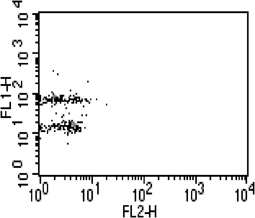
[0067] Embodiment 1, leukemia cell NB 4 Binary Flow Cytometry Liquid Array Detection Method of Fusion Protein PML-RARα

[0068] (1) Collection of mononuclear cells in normal human blood: Take a fresh anticoagulated blood sample, mix it with HBSS's solution 1:1, carefully add it to the liquid surface of the cell separation solution, and centrifuge at 1500 rpm for 15 minutes At this time, the cells in the centrifuge tube were divided into four layers from top to bottom, and the second layer of cells was collected, and the cell pellet was repeatedly washed twice with HBSS's solution to obtain the desired cells.

[0069] (2) Leukemia cells NB 4 Extraction of internal fusion protein PML-RARα: NB 4 Cells, K562 cells, HL-60 cells and normal human mononuclear cells were collected, counted, washed with cold PBS buffer once, centrifuged at 2000 rpm for 3 minutes, and the supernatant was removed to collect the cells; NB 4 Cells were mixed with normal human mononuclear cells collected ...

Embodiment 2

[0078] Embodiment 2. The microspheres and the fluorescent codes of the microspheres in this embodiment are the same as those in Embodiment 1.

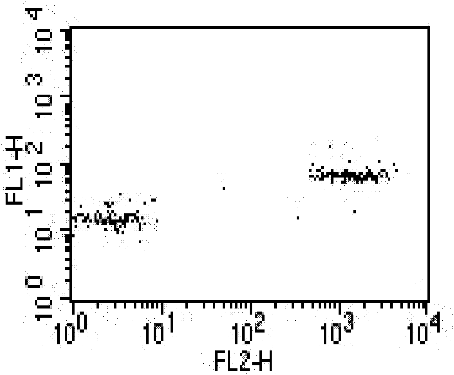
[0079] The binary flow cytometry liquid phase array detection method of leukemia cell K562 fusion protein BCR-ABL, the steps are as follows:

[0080] (1) Collection of mononuclear cells in normal human blood: Take a fresh anticoagulated blood sample, mix it with HBSS's solution 1:1, carefully add it to the liquid surface of the cell separation solution, and centrifuge at 1500 rpm for 15 minutes At this time, the cells in the centrifuge tube are divided into four layers from top to bottom. The second layer of cells was collected, and the cell pellet was repeatedly washed twice with HBSS's solution to obtain the desired cells.

[0081] (2) Extraction of fusion protein BCR-ABL in leukemia cells K562: K562 cells, NB 4 Cells, HL-60 cells and normal human mononuclear cells were collected, counted, washed once with cold PBS buffer, centrifu...


PUM

PropertyMeasurementUnit
diameteraaaaaaaaaa
diameteraaaaaaaaaa

Abstract

The invention relates to a dimensional flow liquid phase array detection method of fusion protein in leukemia cells, characterized in that: a microballoon 1 fluorescently labeled by FITC or Alexa Fluor 488 coated by fusion protein capture antibody and a microballoon 2 fluorescently labeled by FITC or Alexa Fluor 488 with another conentration coated by fusion protein capture antibody form a dimensional flow liquid phase array for detecting fusion protein in leukemia cells, after the co-incubation of the microballoon 1, the microballoon 2 , the fusion protein, a reported antibody and / or a secondary antibody fluorescently labeled by PE, a flow cytometry is used for detecting the dimensional flow liquid phase array, and the fusion protein expression value is expressed as a ratio of the PE fluorescence intensity of the microballoon 1 to the PE fluorescence intensity of the microballoon 2. The method can be applied in the biomedicine research fields, such as leukemia pathology, molecular diagnosis, and discovery of anti-leukemic medicines. The method has the advantages of effective elimination of system error, rapidness, and accuracy.

Description

technical field [0001] The invention belongs to the field of biotechnology, and in particular relates to a method for detecting fusion proteins in leukemia cells. Background technique [0002] Flow liquid array (also known as flow microsphere array) is a molecular detection platform that can guarantee information quality and provide relatively high throughput. The liquid chip technology with the same technical principle is the first clinical array analysis approved by the US FDA in 2001. technology. In the flow liquid phase array, microsphere-anchored capture molecules (antibodies or nucleic acid probes) capture target molecules, fluorescently labeled reporter molecules (antibodies or nucleic acid probes) report target molecules, one or several different concentrations Arrays of fluorescently encoded microspheres can be used for multiparameter parallel analysis. Array decoding commonly used biomedical engineering instrument flow cytometer or special liquid chip instrument....

Claims


Application Information

Patent Timeline
Patent Type & Authority Patents(China)
IPC IPC(8): G01N33/68G01N21/64
Inventor 兰文军王海燕王哲理闫磊
Owner 山东泉力生物科技有限公司
Why Eureka
Try Eureka