Force tracing method for atomic force microscope (AFM)

An atomic force microscope and tracer technology, used in scanning probe microscopy, measuring devices, instruments, etc.

Active Publication Date: 2012-09-12
CHANGCHUN INST OF APPLIED CHEMISTRY - CHINESE ACAD OF SCI
View PDF5 Cites 15 Cited by
  • Summary
  • Abstract
  • Description
  • Claims
  • Application Information

AI Technical Summary

Problems solved by technology

[0006] In order to solve the problem that the force signal detected by AFM single-molecule force spectroscopy in the prior art is easily considered to be generated by the movement of the AFM probe itself rather than the dynamic process on the cell surface, the present invention provides a force display for an atomic force microscope tracking method

Method used


Image

  • Force tracing method for atomic force microscope (AFM)
  • Force tracing method for atomic force microscope (AFM)
  • Force tracing method for atomic force microscope (AFM)

Examples


Embodiment 1

[0034] Example 1 Study on the Dynamic Process of Glucose Molecule Transport on the Cell Membrane

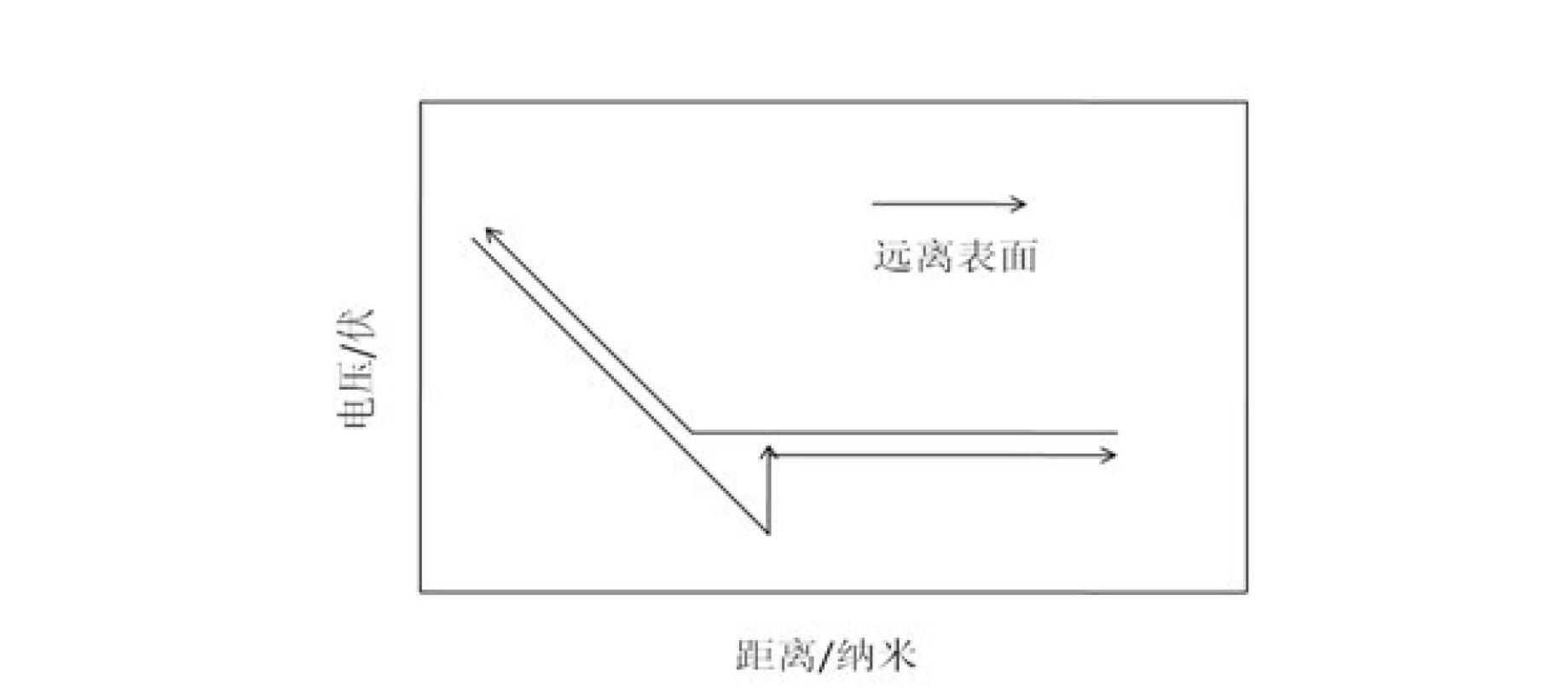
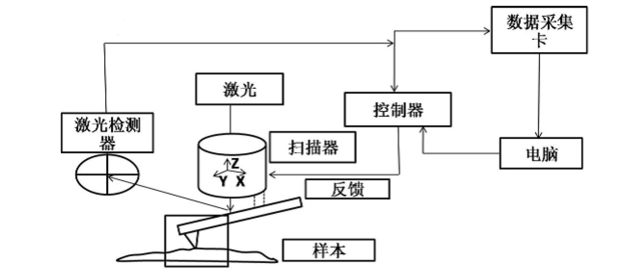
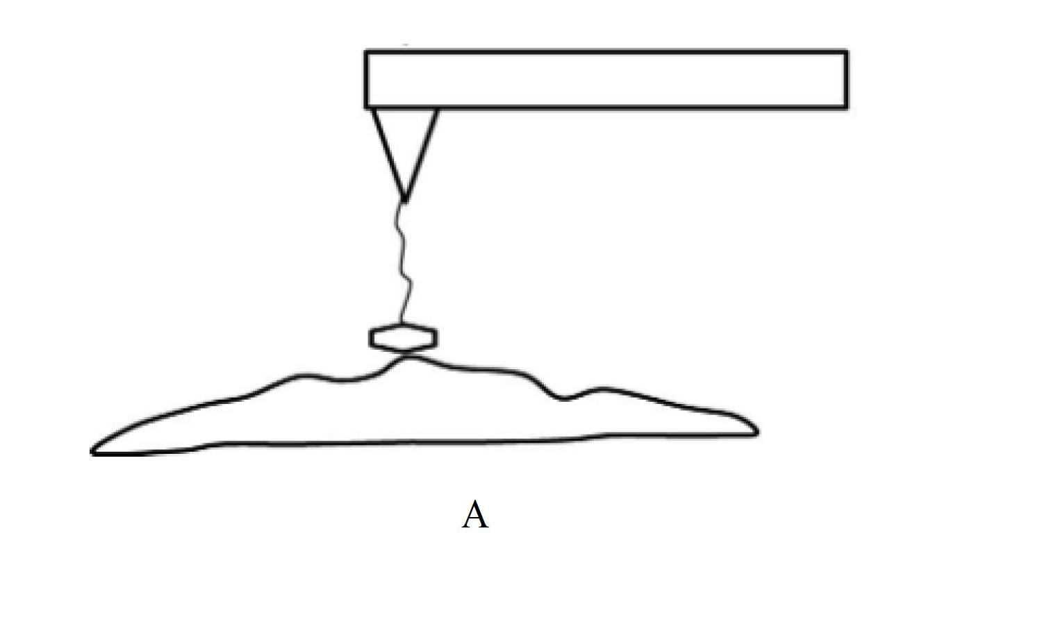
[0035] After the needle is inserted under the control of the feedback regulation system, the position of the AFM probe in the Z direction when the AFM probe is in contact with the cell membrane surface is determined through the obtained force-distance curve; the glucose molecule is modified on the AFM probe tip, and the setting The force when the AFM probe is in contact with the cell membrane surface is within 100 piconewtons. Turn on the software adjustment system to make the AFM probe approach the cell membrane surface gradually. , the microcantilever deflects immediately; use the data acquisition card to collect the relationship of the microcantilever deflection with time and input it to the computer for analysis and processing.

[0036] Such as Figure 4 In A, B, C and Figure 5 As shown in A and B, the experimental results show that the advantages of the present invention ...

Embodiment 2

[0037] Example 2 Research on the Dynamic Process of Endocytosis of Gold Nanoparticles in Cell Membrane

[0038] After the needle is inserted under the control of the feedback regulation system, the position of the AFM probe in the Z direction when the AFM probe is in contact with the cell membrane surface is determined through the obtained force-distance curve; the gold nanoparticles are modified on the AFM probe tip, and the Set the force when the AFM probe is in contact with the cell membrane surface within 100 piconewtons, turn on the software adjustment system, and make the AFM probe gradually approach the cell membrane surface. Needle, the micro-cantilever deflects immediately; use the data acquisition card to collect the relationship of the micro-cantilever deflection with time and input it to the computer for analysis and processing.

[0039] Such as Figure 6As shown, the experimental results show that the advantages of the present invention are: (1) The obtained tran...

Embodiment 3

[0040] Example 3 Research on the Dynamic Process of Amino Acid Molecule Transport on the Cell Membrane

[0041] After the needle is inserted under the control of the feedback regulation system, the position of the AFM probe in the Z direction when the AFM probe is in contact with the cell membrane surface is determined by the obtained force-distance curve; the amino acid molecule is modified on the AFM probe tip, and the setting The force when the AFM probe is in contact with the cell membrane surface is within 100 piconewtons. Turn on the software adjustment system to make the AFM probe approach the cell membrane surface gradually. , the microcantilever deflects immediately; use the data acquisition card to collect the relationship of the microcantilever deflection with time and input it to the computer for analysis and processing.

[0042] Such as Figure 7 As shown, the experimental results show that the advantages of the present invention are: (1) The obtained transport f...


PUM


Abstract

The invention discloses a force tracing method for an atomic force microscope (AFM), which relates to the technical field of micro force measurement methods and solves the problem that force signals obtained by the traditional AFM single molecule force spectrum method are easily regarded as signals generated by motions of an AFM probe but not signals generated in a dynamic process on cell surfaces. The force tracing method for the AFM comprises the following steps of: after needle feed is finished under the control of a feedback regulation system, closing the feedback regulation system, and determining a position of the AFM probe in a Z direction when the AFM probe is contacted with the surface of a tested object through an obtained force-distance curve; gradually approaching the AFM probe to the surface of the tested object, and when the AFM probe is just contacted with the surface of the tested object, closing the feedback regulation system and stopping feeding the probe to ensure that a cantilever is deflected immediately; and acquiring a relation of the deflection of the cantilever along with time by utilizing a data acquisition card. According to the force tracing method, influences of the motions in the Z direction of the AFM probe driven by a piezoelectric ceramic scanner on dynamic process researches of the surface of the tested object are avoided.

Description

technical field [0001] The invention relates to the technical field of microscopic force measurement methods, in particular to a force tracing method of an atomic force microscope. Background technique [0002] The study of the interaction mechanism between biomolecules at the single-molecule level is of great significance for the in-depth understanding of the specific recognition of biomolecules, biochemical processes, and the relationship between molecular structure and function, and has become an important research field in the interdisciplinary fields of biology, chemistry, and physics. A Frontier Direction ([1] Yizhang Chen, Qisho Lin, Single Molecule Behavior and Intracellular Real-Time Detection in Life Sciences, 1, Beijing: Science Press, 2005, 1-11. [2] M.J.J.A.Dvorak, The application of atomic force microscopy to the study of living vertebral cells in culture, Methods, 2003(29): 86-96). [0003] With its unique advantages, AFM single-molecule force spectroscopy pl...

Claims


Application Information

Patent Timeline
Patent Type & Authority Applications(China)
IPC IPC(8): G01Q60/24G01Q60/26
Inventor 王宏达潘延刚蒋俊光单玉萍蔡明军
Owner CHANGCHUN INST OF APPLIED CHEMISTRY - CHINESE ACAD OF SCI
Why Eureka
Try Eureka