Method for detecting rubella virus, quantum dot-labeled immunochromatographic test paper and preparation method thereof

A technology of immunochromatography test strips and quantum dots, which is applied in the field of medical immunoassays, can solve the problems of low accuracy and low sensitivity, and achieve the effects of narrow emission peaks, good luminescence stability, and symmetrical peak shapes

Active Publication Date: 2015-07-22
魏县聚邦新材料科技有限公司
View PDF4 Cites 0 Cited by
  • Summary
  • Abstract
  • Description
  • Claims
  • Application Information

AI Technical Summary

Problems solved by technology

[0003] At present, the commonly used detection method is the colloidal gold method. Although this method is fast, simple and easy to operate, it has low accuracy and low sensitivity.

Method used


Image

  • Method for detecting rubella virus, quantum dot-labeled immunochromatographic test paper and preparation method thereof
  • Method for detecting rubella virus, quantum dot-labeled immunochromatographic test paper and preparation method thereof

Examples


Embodiment 1

[0032] Embodiment 1: A kind of quantum dot-labeled immunochromatographic test paper is provided with plastic plate, nitrocellulose membrane, glass cellulose membrane A, quantum dot-labeled rubella virus IgG monoclonal antibody glass cellulose membrane B, absorbent paper, so The above-mentioned glass cellulose film A is the glass cellulose film purchased on the market without sample;

[0033] Wherein, glass cellulose membrane A, glass cellulose membrane B of quantum dot-labeled rubella virus IgG monoclonal antibody, nitrocellulose membrane, and absorbent paper are pasted sequentially on the plastic plate;

[0034] Wherein, there are rubella virus polyclonal antibody and rabbit anti-mouse secondary antibody at one end of the nitrocellulose membrane, so as to form detection zone T and quality control zone C;

[0035] Wherein, the rubella virus IgG monoclonal antibody labeled with quantum dots is located at the other end of the glass cellulose membrane B, corresponding to the dete...

Embodiment 2

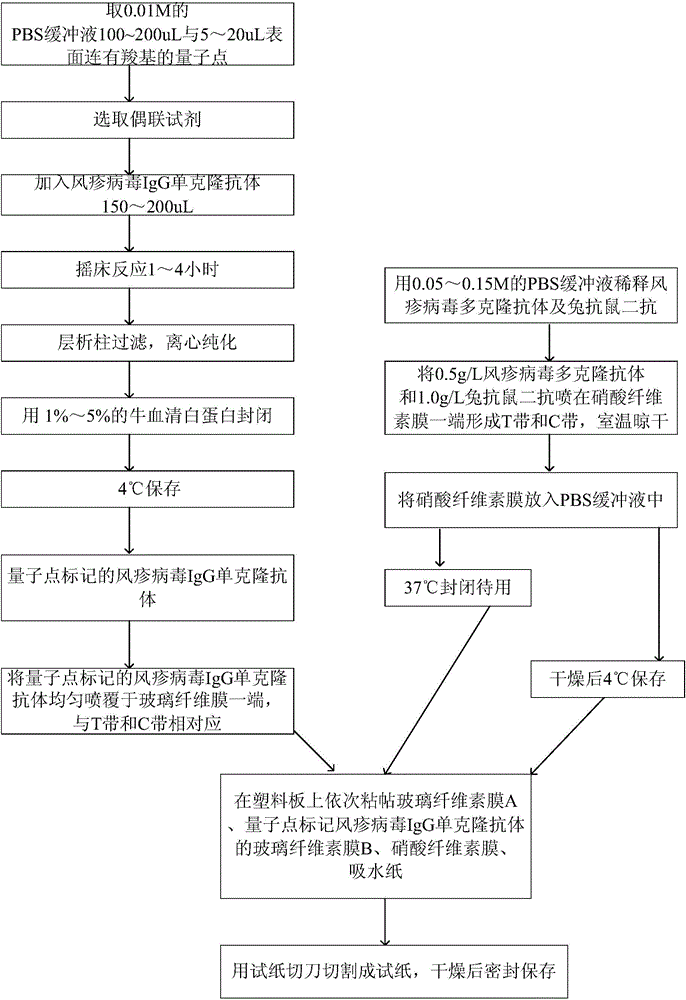
[0041] Embodiment 2: the preparation method of test paper as mentioned above, as figure 1 shown, including the following steps:

[0042] (1) Coupling of quantum dots and rubella virus IgG monoclonal antibody:

[0043] Take 100-200uL of 0.01M PBS buffer and 5-20uL of quantum dots with carboxyl groups on the surface;

[0044] A coupling reagent is selected, and the coupling reagent is selected from hydroxysulfosuccinic acid imide, 1-(3-dimethylaminopropyl)-3 ethylcarbodiamine hydrochloride;

[0045] Add rubella virus IgG monoclonal antibody 150-200uL;

[0046] Shaker reaction for 1 to 4 hours;

[0047] Column filtration, centrifugal purification;

[0048] Block with 1% to 5% bovine serum albumin;

[0049] Store at 4°C;

[0050] (2) Preparation of test paper:

[0051] Dilute rubella virus polyclonal antibody and rabbit anti-mouse secondary antibody with 0.05-0.15M PBS buffer, spray 0.5g / L rubella virus polyclonal antibody and 1.0g / L rabbit anti-mouse secondary antibody on ...

Embodiment 3

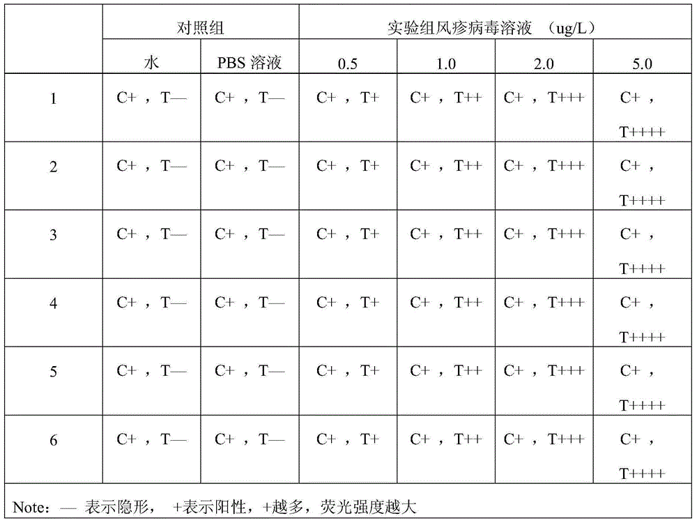
[0058]Embodiment 3: detect rubella virus with described test paper, comprise the following steps: spot sample on the assembled test paper close to one end of rubella virus IgG monoclonal antibody, after reacting for 5min, observe the result in the ultraviolet analyzer . PBS buffer solution and normal human blood were used as blank controls.

[0059] Result judgment: under the premise that the C band shows a red fluorescent band, the intensity of the fluorescent band of the T band is visually compared with the blank. The weaker the fluorescence, the lower the concentration of the tested substance in the test solution.


PUM

PropertyMeasurementUnit
diameteraaaaaaaaaa
concentrationaaaaaaaaaa

Abstract

The invention relates to a medimmune inspection method, and particularly relates to quantum dot-labeled immunochromatographic test paper, and a method for detecting rubella virus by adopting an immunological method. According to the quantum dot-labeled immunochromatographic test paper, a glass cellulose membrane A, a quantum dot-labeled rubella virus IgG monoclonal antibody glass cellulose membrane B, a cellulose nitrate membrane and absorbent paper are sequentially bonded on a plastic board from bottom to top, wherein a rubella virus polyclonal antibody and a rabbit-anti-mouse secondary antibody are arranged on one end of the cellulose nitrate membrane so as to form an inspection strip T and a quality control strip C; a quantum dot-labeled rubella virus IgG monoclonal antibody is located at the other end of the glass cellulose membrane B and corresponds to the inspection strip T and the quality control strip C, and the quantum dot-labeled rubella virus IgG monoclonal antibody is located at one end of a sample feeding point. The inspection sensitivity of the method is higher than of the currently used method by about 1000 times.

Description

technical field [0001] The invention relates to a medical immunological detection method, in particular to a method for immunologically detecting rubella virus by using quantum dot-labeled immunochromatographic test paper. Background technique [0002] Rubella virus is an RNA virus belonging to the Togavirus genus. Rubella virus polyclonal antibody structure is quite stable, there is only one polyclonal antibody type, no subtype. It only infects humans and can grow in rabbit kidney, suckling vole kidney and green monkey kidney cells. The shape is rough and spherical, with a diameter of 50-70nm. It consists of a single-stranded RNA genome and a lipid shell, which contains an electron-dense core and is covered with two layers of loose coats. The virus is heat-labile and quickly loses its activity at 37°C and room temperature. It is cold-resistant and can be stored for a short period of time at -20°C and relatively stable for several months at -60°C. Viability outside the hu...

Claims


Application Information

Patent Timeline
Patent Type & Authority Patents(China)
IPC IPC(8): G01N33/577
CPCG01N33/577G01N2333/19
Inventor 文德敏申有长于晓永
Owner 魏县聚邦新材料科技有限公司
Why Eureka
Try Eureka