Method and device for segmenting three-dimensional breast images

A three-dimensional image, two-dimensional image technology, applied in the direction of image analysis, image data processing, instruments, etc., can solve the problems of low efficiency, complicated semi-automatic segmentation methods, etc., to achieve simple results

Active Publication Date: 2016-03-02
SHANGHAI UNITED IMAGING HEALTHCARE
View PDF5 Cites 0 Cited by
  • Summary
  • Abstract
  • Description
  • Claims
  • Application Information

AI Technical Summary

Problems solved by technology

[0006] What the present invention aims to solve is the problem that manual and user-assisted semi-automatic segmentation methods are relatively complicated and inefficient in the existing three-dimensional breast image segmentation methods

Method used


Image

  • Method and device for segmenting three-dimensional breast images
  • Method and device for segmenting three-dimensional breast images
  • Method and device for segmenting three-dimensional breast images

Examples


Embodiment Construction

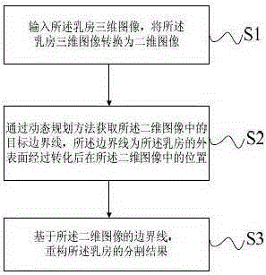
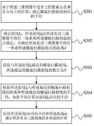
[0035] The present invention will be described in detail below in conjunction with the accompanying drawings and embodiments. The segmentation method of breast three-dimensional image of the present invention is as figure 1 As shown, first, step S1 is performed to input the three-dimensional image of the breast, and convert the three-dimensional image of the breast into a two-dimensional image. Specifically, the three-dimensional image of the breast is converted into a two-dimensional image by the helical scanning method and the polar coordinate method (such as figure 2 shown), the process can be found in Jiahui W., Roger E., and QiangL.,"Segmentationofpulmonarynodulesinthree-dimensionalCTimagesby use of aspiral-scanning technique," Med. Phys. 34(12), 4678-4689 (2007) . The three-dimensional image of the breast obtains a certain number of helical scanning rays in a certain order through the helical scanning method. In this embodiment, the number of helical scanning ray...


PUM


Abstract

The invention provides a method for dividing a breast three-dimensional image and a device thereof. The method for dividing a breast three-dimensional image comprises a step of inputting the breast three-dimensional image and converting the breast three-dimensional image into a two-dimensional image, a step of obtaining the object boundary line of two-dimensional image by a dynamic programming method, and a step of reconstructing the division result of a breast based on the object boundary line of two-dimensional image, wherein the boundary line is the position of the breast in the two-dimensional image after the outer surface of the breast is converted. According to the scheme, the gray, gradient and shape features of the breast three-dimensional image are combined, and the automatic three-dimensional division is accurately and effectively realized.

Description

technical field [0001] The invention relates to the field of image segmentation, in particular to a method and device for segmenting three-dimensional breast images. Background technique [0002] Breast cancer is the most common malignant tumor in middle-aged and elderly women, and its morbidity and mortality are increasing year by year. The curative effect of breast cancer depends more on the stage of the disease, especially in early breast cancer, the cure rate of ductal carcinoma in situ can reach 95%. Therefore, the early detection and diagnosis of breast cancer can effectively improve the cure rate of breast cancer. In clinical practice, magnetic resonance images have been widely used in risk prediction and early diagnosis of breast cancer, and computer-aided diagnosis system helps to improve the accuracy and efficiency of radiologists in reading 3D magnetic resonance images. In the computer-aided diagnosis system based on 3D magnetic resonance images, the segmentation...

Claims


Application Information

Patent Timeline
Patent Type & Authority Patents(China)
IPC IPC(8): G06T7/00
Inventor 姜娈连燕云李强
Owner SHANGHAI UNITED IMAGING HEALTHCARE
Why Eureka
Try Eureka