Method, device and system for labeling medical images

A technology for medical images and images, applied in the field of labeling medical images, can solve the problems of reducing labeling efficiency, lack of a unified collaboration mechanism, and inability to share labeling results, to simplify the acquisition process and improve labeling efficiency.

Inactive Publication Date: 2015-03-25
SIEMENS AG
View PDF6 Cites 42 Cited by
  • Summary
  • Abstract
  • Description
  • Claims
  • Application Information

AI Technical Summary

Problems solved by technology

[0004] However, these existing medical image annotation tools all regard their respective annotation tasks as separate tasks, and there is no unified cooperation mechanis

Method used


Image

  • Method, device and system for labeling medical images
  • Method, device and system for labeling medical images
  • Method, device and system for labeling medical images

Examples


Embodiment Construction

[0048] In order to make the technical solutions and advantages of the present invention clearer, the present invention will be further described in detail below in conjunction with the accompanying drawings and embodiments. It should be understood that the specific embodiments described here are only used to illustrate the present invention, and are not intended to limit the protection scope of the present invention.

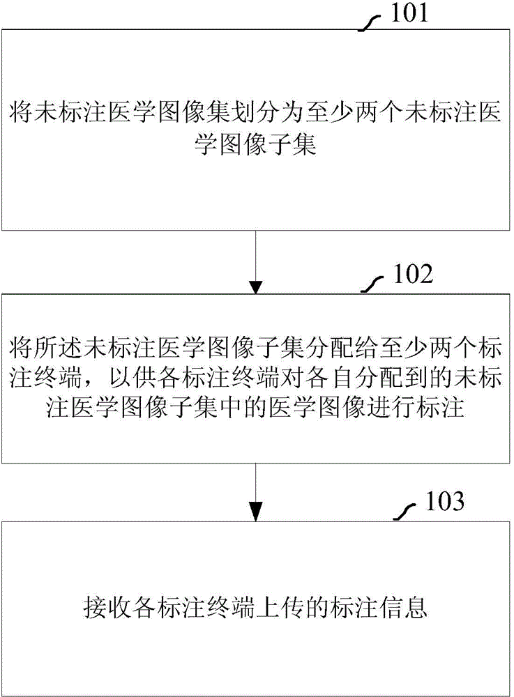
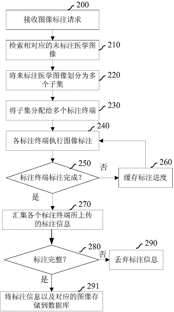
[0049] figure 1 It is a flowchart of a method for labeling medical images according to an embodiment of the present invention. Such as figure 1 As shown, the method includes:

[0050] Step 101: Divide the unlabeled medical image set into at least two unlabeled medical image subsets.

[0051] The medical image database can be established in advance on the network side. An unlabeled medical image set and an annotated medical image set are stored in the medical image database. The labeled medical image set contains medical images that have been labeled; the un...


PUM


Abstract

The embodiment of the invention provides a method for labeling medical images. The method includes the steps of dividing an unlabeled medical image set into at least two unlabeled medical image subsets, distributing the unlabeled medical image subsets to at least two labeling terminals so that each labeling terminal can label the medical images in the corresponding distributed unlabeled medical image subset, and receiving labeling information unloaded by each labeling terminal. Accordingly, the embodiment of the invention further provides a device and system for labeling the medical images. Through the method, device and system, the coordinative medical image labeling solution can be obtained, and the labeling efficiency is improved.

Description

technical field [0001] The present invention relates to the technical field of image processing, in particular to a method, device and system for labeling medical images. Background technique [0002] Medical images produced by various radiological modalities contain a large amount of information such as anatomy and pathology, which can provide a large amount of visual information, and constitute with non-image data such as genomics, proteomics, histopathology, and clinical laboratory data. complementary. In the process of medical image processing, it is often necessary to give precise annotations to the boundaries of regions of interest (ROI). However, manually annotating medical images is usually time-consuming and expensive, and the annotation results vary from person to person. In addition, the user has to spend dedicated time and space to perform labeling, and it is also difficult to share the labeling results with others. [0003] Existing medical image annotation t...

Claims


Application Information

Patent Timeline
IPC IPC(8): G06F19/00
Inventor 郑杰
Owner SIEMENS AG
Why Eureka
Try Eureka