
A preparation method of 3D printing orthopedic fixation device

A 3D printing, orthopedic technology, applied in fixator, medical science, surgery, etc., can solve problems such as inability to cooperate, and achieve the effect of accurate contact surface, high manufacturing precision and low price

Inactive Publication Date: 2017-03-15
黄若景 +1
View PDF7 Cites 0 Cited by
  • Summary
  • Abstract
  • Description
  • Claims
  • Application Information

AI Technical Summary

Problems solved by technology

[0002] Colles fracture: dorsally comminuted, dorsally angulated and dorsally displaced extraarticular fractures of the distal radius with shortening of the radius, orthopedic fixation devices for Colles fractures made by traditional industrial methods: volar distal radius locking plate screw system , and the steel plate is mass-produced, because each patient’s fracture position is different, and the radius structure is also different, so the steel plate cannot perfectly match the patient’s radius when used.

Method used


Image

  • A preparation method of 3D printing orthopedic fixation device
  • A preparation method of 3D printing orthopedic fixation device

Examples


Embodiment Construction

[0025] The preferred embodiments of the present invention will be described in detail below in conjunction with the accompanying drawings, so that the advantages and features of the present invention can be more easily understood by those skilled in the art, so as to define the protection scope of the present invention more clearly.

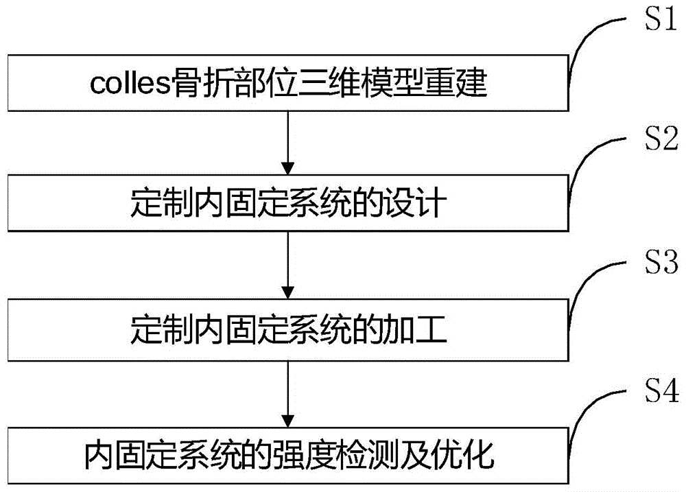
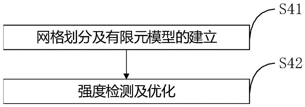
[0026] refer to figure 1 and figure 2 As shown, the present invention provides a method for preparing a 3D printed orthopedic fixation device, comprising the following steps,

[0027] S1, 3D model reconstruction of colles fracture site (take the orthopedic orthopedic fixation device for this kind of fracture as an example),

[0028] The three-dimensional model of the radius is reconstructed according to the two-dimensional images of the patient's preoperative CT and MRI scans;

[0029] S2. Design of customized orthopedic fixation system (it can be either orthopedic fixation device or external fixation device),

[0030] Compared with the norma...


PUM


Abstract

The invention discloses a preparation method of a 3D (three-dimensional) printing orthopaedic fixation device. The method comprises the following steps of S1, rebuilding a three-dimensional model for a colles fracture part; S2, designing a customized orthopaedic fixation system; S3, machining the customized orthopaedic fixation system; S4, detecting and optimizing the strength of the material of an orthopaedic fixation instrument. The size and the shape of the orthopaedic fixation system can be customized according to the shape of skeleton and a soft tissue of a patient, so that the individual customization of the orthopaedic fixation system is realized, the design of the orthopaedic fixation system can be flexibly changed according to the positions and sizes of bone fractures; the surface between the orthopaedic fixation system and a bone is precise, and the principle of stress dispersion is met; printing is performed by the fixer through 3D printing technology, so that the manufacturing precision is high, the price is low, and the fixer is suitable of individual customization; according to the manufactured orthopaedic fixation device, after strength detection and optimized design, the problem that the fixer and the bone or the soft tissue are not engraved or the problem of stress difference between the fixer and the bone or the soft tissue are further solved.

Description

technical field [0001] The invention relates to the technical field of colles fracture reconstruction, and more specifically, relates to a preparation method of a 3D printed orthopedic fixation device. Background technique [0002] Colles fracture: dorsally comminuted distal radius fracture, dorsally angulated and dorsally displaced extra-articular fracture with radius shortening, orthopedic fixation device for Colles fracture made by traditional industrial methods: distal radius volar locking plate screw system , while the steel plate is mass-produced, because each patient's fracture location is different, and the radius structure is also different, so the steel plate cannot perfectly fit the patient's radius when used. Contents of the invention [0003] The purpose of the present invention is to provide a method for preparing a 3D printed orthopedic fixation device that can perfectly match the fracture of each patient, has good curative effect, and low medical expenses. ...

Claims


Application Information

Patent Timeline
Patent Type & Authority Patents(China)
IPC IPC(8): A61B17/58B33Y10/00B33Y50/00
CPCA61B17/58A61B2017/00526A61B2017/568
Inventor 黄若景关宏刚陈志维曹燕明张美超丁玲黄银凤
Owner 黄若景
Learn More