Formula and preparation method of photocuring 3D printing haversian system artificial bone scaffold

A 3D printing and light-curing technology, applied in the medical field, can solve the problems of limited shape, difficult to control pore size and interconnection degree, solvent residue, etc., and achieve the effect of easy implementation and promotion, simple and convenient preparation process, and promotion of bone repair process

Inactive Publication Date: 2019-07-23
FOURTH MILITARY MEDICAL UNIVERSITY
View PDF6 Cites 3 Cited by
  • Summary
  • Abstract
  • Description
  • Claims
  • Application Information

AI Technical Summary

Problems solved by technology

[0004] The technical problem to be solved by the present invention is to provide the formulation and preparation method of photocured 3D printed Haversian system artificial bone scaffold, so as to solve the problem that the traditional tissue engineering scaffold preparation method is difficult to control the pore size and interconnection degree, the preparation shape is limited, and there is no solvent. Disadvantages such as residual

Method used


Image

  • Formula and preparation method of photocuring 3D printing haversian system artificial bone scaffold
  • Formula and preparation method of photocuring 3D printing haversian system artificial bone scaffold
  • Formula and preparation method of photocuring 3D printing haversian system artificial bone scaffold

Examples


Embodiment 1

[0035] The formulation of the light-curing 3D printing Haversian system artificial bone scaffold listed in this example is made of the following raw materials:

[0036] Collagen (medical grade) 5%, normal saline 10%, nano-hydroxyapatite (medical grade) 43%, photosensitive resin (medical grade) 40%, photoinitiator 1%, dispersant 1%.

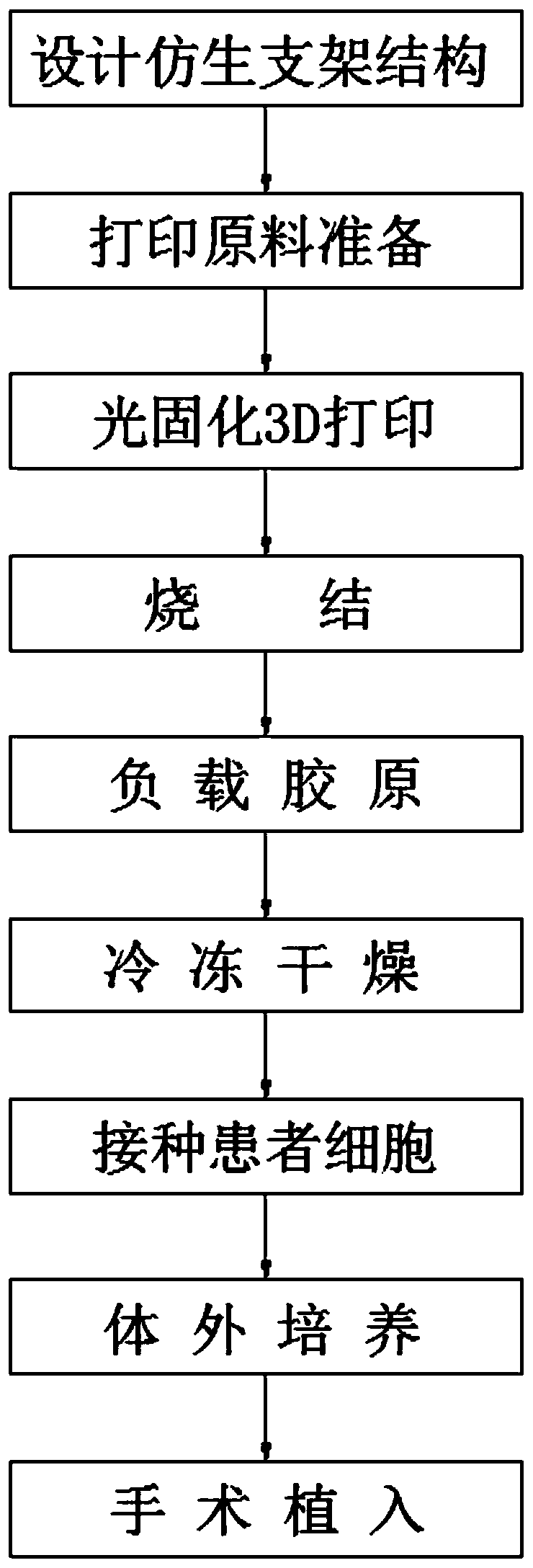
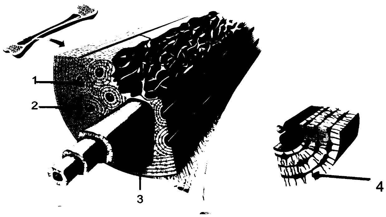
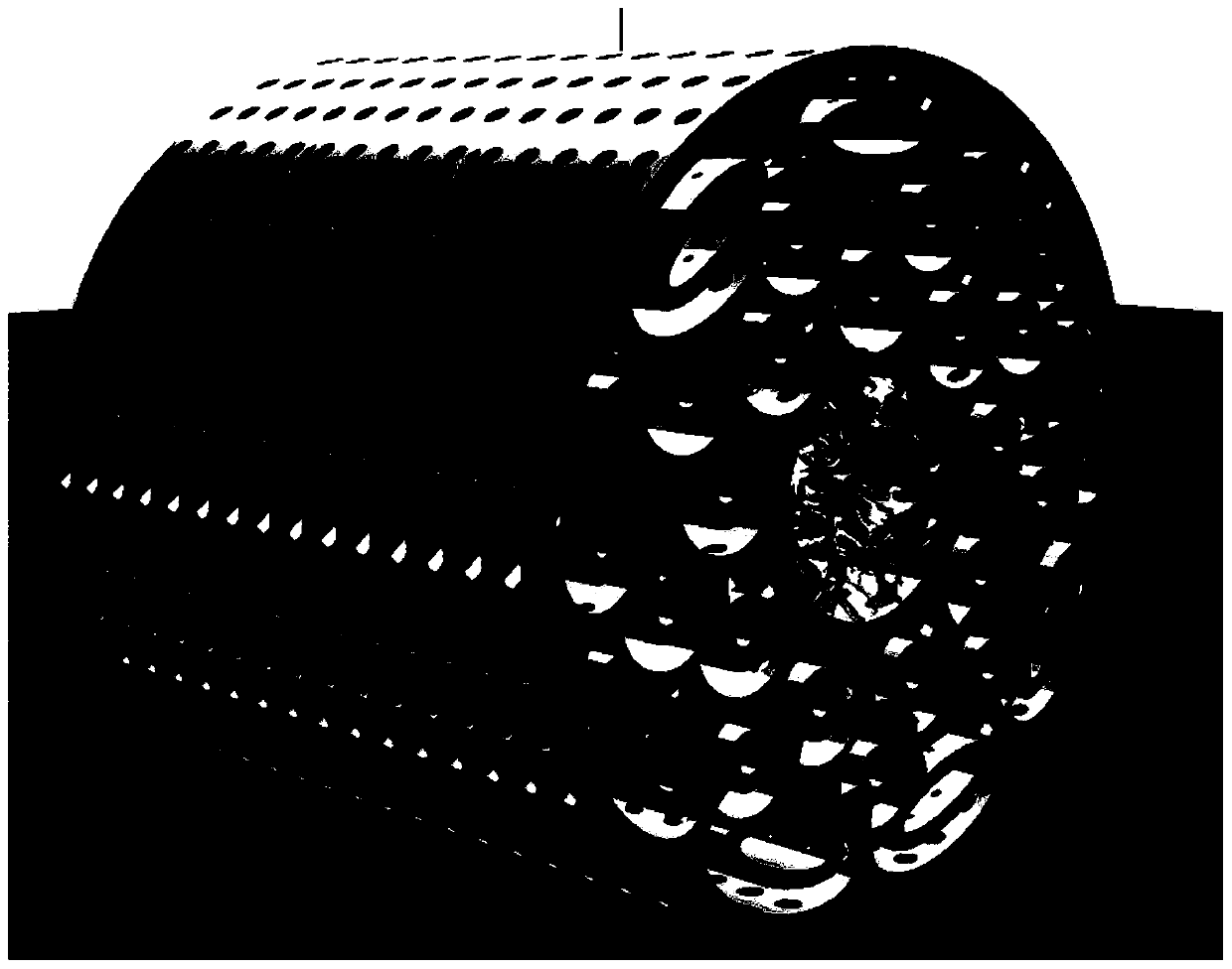
[0037]It is made according to the following method. Firstly, micro-CT scans the human bone metaphysis specimen (such as human distal femur or proximal tibia), obtains micro-CT data of human trabecular bone, reconstructs the structure of trabecular bone in 3D, and saves it as an STL file. , combining the reconstructed bone trabecular structure with the scaffold designed by the bionic Harvard tube system, that is, the central part is the bone trabecular structure, and the peripheral part is the Haversian tube system structure, and the STL file of the bionic artificial bone scaffold is obtained, and the nano-hydroxyapatite Stone (medical grade), phot...

Embodiment 2

[0040] The formulation of the light-curing 3D printing Haversian system artificial bone scaffold listed in this example is made of the following raw materials:

[0041] Collagen (medical grade) 3.5%, normal saline 7.5%, nano-hydroxyapatite (medical grade) 56.25%, photosensitive resin (medical grade) 30%, photoinitiator 0.75%, dispersant 2%.

[0042] It is made according to the following method. Firstly, micro-CT scans the human bone metaphysis specimen (such as human distal femur or proximal tibia), obtains micro-CT data of human trabecular bone, reconstructs the structure of trabecular bone in 3D, and saves it as an STL file. , combining the reconstructed bone trabecular structure with the scaffold designed by the bionic Harvard tube system, that is, the central part is the bone trabecular structure, and the peripheral part is the Haversian tube system structure, and the STL file of the bionic artificial bone scaffold is obtained, and the nano-hydroxyapatite Stone (medical gr...

Embodiment 3

[0045] The formulation of the light-curing 3D printing Haversian system artificial bone scaffold listed in this example is made of the following raw materials:

[0046] Collagen (medical grade) 5%, physiological saline 10%, nano-hydroxyapatite (medical grade) 61.5%, photosensitive resin (medical grade) 20%, photoinitiator 0.5%, dispersant 3%.

[0047] It is made according to the following method. Firstly, micro-CT scans the human bone metaphysis specimen (such as human distal femur or proximal tibia), obtains micro-CT data of human trabecular bone, reconstructs the structure of trabecular bone in 3D, and saves it as an STL file. , combining the reconstructed bone trabecular structure with the scaffold designed by the bionic Harvard tube system, that is, the central part is the bone trabecular structure, and the peripheral part is the Haversian tube system structure, and the STL file of the bionic artificial bone scaffold is obtained, and the nano-hydroxyapatite Stone (medical ...


PUM


Abstract

The invention discloses a formula and a preparation method of a photocuring 3D printing haversian system artificial bone scaffold. The scaffold is prepared from the following raw materials: 2-5% of collagen (medical grade), 5-10% of normal saline, 43-61.5% of nano hydroxyapatite (medical grade), 20-40% of photosensitive resin (medical grade), 0.5-1% of photoinitiator and 1-3% of dispersant, wherein the sum of the weight percentages of the components is 100%. The preparation method of the photocuring 3D printing haversian system artificial bone scaffold is inspired by natural bone microstructure, uses a bionic method for designing an artificial bone internal pore structure, and promotes the vascularization formation in an artificial bone implant body so as to promote the bone repair process. The application of the 3D printing method realizes individualized customization of a bone defect part of a patient, can accurately control the appearance, the internal structure and the degradationrate of the artificial bone, really realizes customization, vascularization and intelligence of the artificial bone scaffold, and provides a new idea for design and development of the artificial bonescaffold.

Description

technical field [0001] The invention relates to the field of medical technology, in particular to a formulation of a light-cured 3D printed Haversian system artificial bone scaffold and a preparation method thereof. Background technique [0002] In the field of orthopedics, the regeneration of large bone defects is a clinical problem that has been difficult to solve so far. Among them, angiogenesis plays an important role in the process of bone regeneration. A large number of basic experiments have proved that the microporous structure can induce endothelial cells to promote the implantation of scaffolds. of vascularization. Traditional three-dimensional scaffolds have the disadvantages of low porosity and low connectivity, which hinder the formation of blood vessels and bone formation inside the scaffold. In recent years, 3D printed tissue engineering scaffolds have developed rapidly in bone tissue engineering. The advantage of 3D printing lies in the ability to precisely ...

Claims


Application Information

Patent Timeline
Patent Type & Authority Applications(China)
IPC IPC(8): A61L27/12A61L27/14A61L27/24A61L27/38A61L27/50A61L27/56A61L27/58B33Y50/00B33Y70/00B33Y80/00
CPCA61L27/12A61L27/14A61L27/24A61L27/3808A61L27/3834A61L27/50A61L27/56A61L27/58A61L2430/02B33Y50/00B33Y70/00B33Y80/00
Inventor 裴国献宋岳毕龙林楷丰程朋真李俊琴吴昊高祎雷星张帅帅
Owner FOURTH MILITARY MEDICAL UNIVERSITY
Why Eureka
Try Eureka