

Device and method for detecting blood vessel
A technology of blood vessels and visible light, which is applied in the fields of application, blood flow measurement, and diagnosis by using light. It can solve the problem of not being able to directly display the surrounding tissue of microvascular, and achieve the effect of low energy, high image quality, and improved contrast.
- Summary
- Abstract
- Description
- Claims
- Application Information
AI Technical Summary
Benefits of technology
Problems solved by technology
Method used
Image


Examples
Embodiment 1
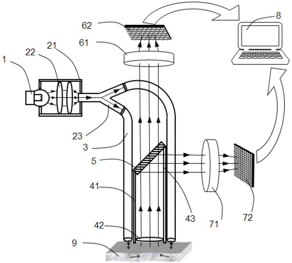
[0041] see figure 1 , the device for detecting blood vessels described in this embodiment includes a visible light source 1, a fiber coupler 2, an illumination fiber 3, an imaging receiving channel 4, an infrared beam splitter 5, a far-infrared lens zoom system 61, a far-infrared Detector 62, visible light lens zoom system 71, visible light image sensor 72, and image processing system 8; the visible light source 1 is used to emit visible light that can be absorbed by red blood cells; the optical fiber coupler 2 is arranged in the advancing direction of the visible light, It is used to receive the visible light emitted by the visible light source 1; the illumination optical fiber 3 is connected to the optical fiber coupler 2, and is used to transmit the visible light, export the visible light and project it onto the surface of human tissue 9; the imaging receiving The channel 4 is used to collect the visible light and the far-infrared radiated from the tissue surface scattered ...
Embodiment 2
[0066] In order to better understand the present invention, the structure of the present invention will be further introduced below in conjunction with a specific embodiment. The contents not described in detail in this embodiment are the same as those in Embodiment 1.
[0067] Such as figure 2 As shown, the device for detecting blood vessels of the present invention includes a visible light source 1, a fiber coupler 2, an illumination fiber 3, an imaging receiving channel 4, an infrared beam splitter 5, a far-infrared lens zoom system 61, a far-infrared detector 62, Visible light lens zoom system 71, visible light image sensor 72 and image processing system 8; the fiber coupler 2 includes a fixed cover 21, a condenser lens 22 and an optical splitter 23, the visible light source 1 is fixed in the fixed cover 21, and the progress of visible light Condenser 22 and optical splitter 23 are installed sequentially in the direction, one end of optical splitter 23 is fixedly connect...
Embodiment 3
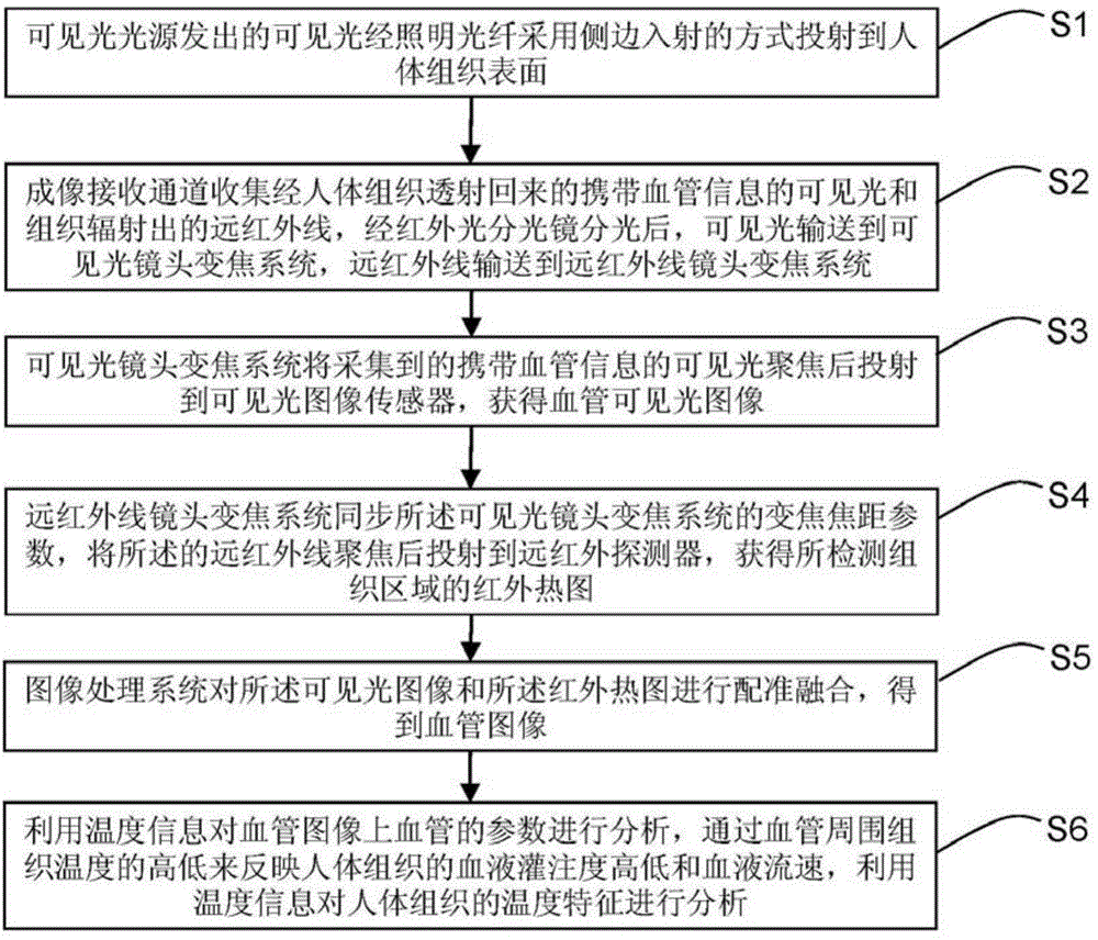
[0078] The invention also provides a method for detecting blood vessels. see image 3 , using the device for detecting blood vessels in the above embodiment 1 or 2 to detect human tissue microvessels, comprising the following steps:
[0079] S1. Visible light emitted by the visible light source is projected onto the surface of human tissue through the illuminating fiber in the way of side incidence;
[0080] S2. The imaging receiving channel collects the visible light carrying microvascular information transmitted back through the human tissue and the far-infrared radiated from the tissue detection area. After being split by the infrared beam splitter, the visible light is sent to the zoom system of the visible light lens, and the far-infrared light is sent to the far-infrared lens. zoom system;
[0081] S3. The visible light lens zoom system focuses the collected visible light carrying microvessel information and projects it to the visible light image sensor to obtain a vis...
PUM
| Property | Measurement | Unit |
|---|---|---|
| Diameter | aaaaa | aaaaa |
| Length | aaaaa | aaaaa |
| Angle of incidence | aaaaa | aaaaa |
Abstract
Description
Claims
Application Information