Dynamic multi-spectral X-ray projection imaging

a multi-spectral x-ray and projection imaging technology, applied in the direction of instruments, patient positioning for diagnostics, applications, etc., can solve the problems of measurement lack of quantitative accuracy, doubts about the likelihood of significant changes in patient outcomes, etc., to improve the conspicuity of lesion and patient outcomes, improve measurement and diagnostic results, and improve the effect of cad algorithms

Inactive Publication Date: 2005-09-27
FOREVISION IMAGING TECH LLC
View PDF34 Cites 202 Cited by
  • Summary
  • Abstract
  • Description
  • Claims
  • Application Information

AI Technical Summary

Benefits of technology

[0025]The instrumentalities described herein overcome the problems outlined above by providing method and apparatus for use in multispectral X-ray imaging applications that may be used to improve measurements and diagnostic results. For example, in medical applications these instrumentalities may improve lesion conspicuity and patient outcomes. Use of co-registered multi-spectral data may improve the efficacy of CAD algorithms in detecting, characterizing, and classifying lung and other lesions. In other applications, threat diagnosis may be improved, for example, by diagnosing that threat-related materials are present, such as a firearm, explosives, shielded nuclear materials, and radiological dispersal devices.
[0026]Using multi-spectral data acquisition in a single scan, as described below, advantageously eliminates motion and mis-registration artifacts between the various multi-spectral projections. Co-registered elimination of bone superposition may bring significant improvements in lesion conspicuity. Use of multiple energies may further improve separation of body constituents and allow better anatomy visualization and lesion characterization, particularly in the presence of contrast medium. As applied to chest imaging, better imaging test accuracy, in conjunction with improved lung cancer treatment and management techniques, may provide better long-term patient outcomes. The rate of false negatives may be improved, as compared to conventional chest radiography. The rate of false positives may also be reduced through improved contrast, conspicuity, and characterization. A reduced rate of false positives may reduce the burden on the patient and the healthcare system, by avoiding costly unnecessary and potentially harmful follow-up and interventions. Similar advantages are obtained in security and industrial applications.
[0027]Systems and methods are described to dynamically and adaptively vary the X-ray spectrum, during a scan of the body, to produce a set of results that differentiate a far wider range of body constituents and improve image contrast in substantially the same examination time as compared to the prior art.
[0035]The wide-band X-ray source may be used in conjunction with detectors that have the capability of count individual photon events and determine the energy of each such X-ray. The systems and methods described below may process the data acquired in T sets of measurements for M spectra and decompose them onto a set of N basis functions representative of body constituents. The processing may, for example, proceed on the basis of spectral and spatial relationships between the T sets of measurements each allocated to a different one of the M spectra, and / or among the T sets of measurements allocated between the M spectra. An ability to dynamically adjust spectral selection in synchrony with scanning excursions is particularly advantageous when used with the wideband source and filtration assembly.
[0037](a) projecting X-ray radiation along paths through a body by scanning a beam of X-ray radiation with respect to the body in synchronicity with system configuration for selective emission of X-ray radiation having M sets of spectral characteristics, the paths extending from an X-ray source to a detector and with an x-ray source excursion and detector configuration allowing acquisition of sufficient data for at least one of a tomographic reconstruction and a tomosynthesis reconstruction;
[0042]The CT imaging method may be implemented using commercially available components that have been assembled to operate as described above when programmed with instructions to implement the decomposition. The selection of M spectra may be facilitated by a wideband source X-ray tube used in combination with a specifically constructed filtration system for selection of a specified one of the M spectral sets. The wide-band X-ray source may be used in conjunction with detectors that have the capability of count individual photon events and determine the energy of each such X-ray. An ability to dynamically adjust spectral selection in synchrony with scanning excursions is particularly advantageous when used with the wideband source and filtration assembly.

Problems solved by technology

The challenge for CAD in chest radiography is to distinguish true nodules from overlapping shadows of ribs and vessels.
In lung imaging, CT has demonstrated significant sensitivity, but its relatively low specificity has raised concerns about the likelihood of significant changes in patient outcomes.
Unfortunately, one problem with system 10 is that the tomographic map of section 24 can be used to identify relatively few constituents of body 16, and measurements lack quantitative accuracy.

Method used


Image

  • Dynamic multi-spectral X-ray projection imaging
  • Dynamic multi-spectral X-ray projection imaging
  • Dynamic multi-spectral X-ray projection imaging

Examples


example 1

Modeling of Beam Spectra Generation and Detection

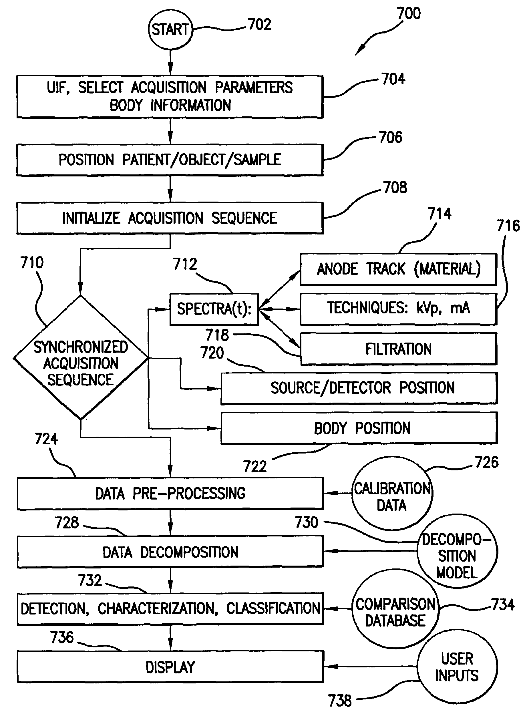
[0194]It is possible to mathematically model X-ray spectra, filtration, phantom attenuation, and detector absorption, for example, including the spectral attenuation equation, as shown by equation (1.3). As will be seen below, this model confirms a physical basis for the feasibility of full-frame sampling and narrow-beam X-ray scanning, for example in lung imaging as well as for other applications described above. The output of this model may be used to provide test input sets for use in further modeling. In particular, key aspects of signal, noise, dose, and spectra separation have been modeled and confirmed by empirical studies.

[0195]In practical X-ray beam imaging, four parameters are available to shape the X-ray spectrum. These are the X-ray target material, the amount and type of filtration used, the kVp applied, and the tube current that is selected. Calculations to provide an X-ray spectra may, for example, be performed using t...

example 2

Single-scan Dual-energy Imaging

[0204]Initial multi-spectral investigations were modeled with a dual-energy scan configuration, using typical spectra as obtained at 120-kVp and 2-mm Al and 3-mm Cu filtration; and 60-kVp and 2-mm Al filtration, respectively. With a beam width of 10-mm at the detector, and a pixel size of 127-μm, the primary beam illuminated 79 pixels and the effective exposure time was 315-ms for any point in the object. Tube currents of 12 and 6-mAs at 120 and 60-kVp respectively led to a (constant) tube current of 60-mA, and 52 and 26 samples at 120 and 60 kVp respectively. Tables 4 and 5 below list various model input parameters and output relating to the phantom, dual-energy techniques, filtration, and detector parameters.

[0205]

TABLE 4Phantom, techniques, and filtration parameters fordual-energy single scan acquisitionBeam widthpixel sizeReadoutExposure Timemmmicrons# of pixelsmsmsTube mAScan LengthScan time1012779 4315.05743013.5kVp:12060mAs:126FilterAl-filt:22Fi...

example 3

Single-scan Imaging with Six Spectra

[0208]A simulated a six-spectra acquisition is shown below for illustration of how this methodology may be applied in a more general sense. Tables 6 and 7 list the corresponding spectral characteristics. In this experiment, two additional low-energy spectra were designed to have mean energies just below and just above the K-edge of Iodine.

[0209]

TABLE 6Techniques for a six-spectra AAPM chest phantom simulationBeam widthpixel sizeReadoutExposureTube currentScan lengthScan timemmmicrons# of pixelsmsmsmAmmseconds12127946566.912043020.3kVp:140120100806040Total Al:Al555455FilterAl-filt:222122FilterCu554100FilterCe0.40FilterHafnium0.2Filteriodine0.1PhantomAcrylic100100100100100100PhantomWater000000PhantomAl333333

[0210]

TABLE 7X-ray characteristics of a six-spectra AAPM-phantom simulationAir exposuresSettings:kVp:140120100806040140-120mAs:571561520(or sum: Σ=)SID-scaled mAs2.63.67.73.17.710.2Fluence / mm*2218134124946134016109091171207318211Kerma (scaled to ...


PUM

PropertyMeasurementUnit
widthaaaaaaaaaa
energyaaaaaaaaaa
sizeaaaaaaaaaa

Abstract

A multispectral X-ray imaging system uses a wideband source and filtration assembly to select for M sets of spectral data. Spectral characteristics may be dynamically adjusted in synchrony with scan excursions where an X-ray source, detector array, or body may be moved relative to one another in acquiring T sets of measurement data. The system may be used in projection imaging and / or CT imaging. Processed image data, such as a CT reconstructed image, may be decomposed onto basis functions for analytical processing of multispectral image data to facilitate computer assisted diagnostics. The system may perform this diagnostic function in medical applications and / or security applications.

Description

RELATED APPLICATIONS[0001]This application claims benefit of priority to provisional application Ser. Nos. 60 / 482,819 filed Jun. 25, 2003, No. 60 / 490,300 filed Jul. 25, 2003, No. 60 / 490,291 filed Jul. 25, 2003, No. 60 / 490,310 filed Jul. 25, 2003, No. 60 / 490,299 filed Jul. 25, 2003, and No. 60 / 490,290 filed Jul. 25, 2003, all of which are hereby incorporated by reference.BACKGROUND[0002]1. Field of the Invention[0003]The invention pertains to dynamic multispectral x-ray imaging of objects, such as an object, animal, tissue sample, or human body.[0004]2. Description of the Related Art[0005]X-rays are generated by a high voltage X-ray tube that is driven by a control unit and interconnected through cabling to a high-voltage generator. A beam of the X-rays pass through a subject body during X-ray examination and onto an X-ray detector array. The X-ray detector array may be a flat panel detector, for example as described in U.S. Pat. No. 6,403,965 issued to Ikeda et al. Other detection d...

Claims


Application Information

Patent Timeline
Patent Type & Authority Patents(United States)
IPC IPC(8): A61B6/03A61B6/00G01N23/00G21K1/00G21K1/12A61B6/02A61B6/04A61B6/06G21K1/10H05G1/60
CPCA61B6/032A61B6/4028A61B6/4035A61B6/4042A61B6/405A61B6/4241A61B6/4488A61B6/482A61B6/488A61B6/541A61B6/542A61B6/563G21K1/10A61B6/508A61B6/027A61B6/0457A61B6/06A61B6/4085A61B6/4275A61B6/4441A61B6/484A61B6/5288A61B6/583Y10S378/901A61B6/0487
Inventor BESSON, GUY M.
Owner FOREVISION IMAGING TECH LLC
Why Eureka
Try Eureka