Eureka AIR delivers breakthrough ideas for toughest innovation challenges, trusted by R&D personnel around the world.

Prostate 3D Image Segmentation Method Based on Gigabit Electron Computed Tomography

An electronic computer and three-dimensional image technology, which is applied in the field of image processing, can solve the problems of ineffective detection of prostate tissue, low image clarity and resolution, and inconspicuous features of the prostate region, so as to reduce time complexity and avoid Iterative phenomenon, outstanding effect

Active Publication Date: 2019-02-22
HANGZHOU VOCATIONAL & TECHN COLLEGE +1
View PDF2 Cites 0 Cited by
  • Summary
  • Abstract
  • Description
  • Claims
  • Application Information

AI Technical Summary

Problems solved by technology

The imaging quality of B-ultrasound images is not high, that is, the clarity and resolution of the images are relatively low, and the characteristics of the prostate area are not obvious, so the prostate tissue cannot be effectively detected

Method used


Image

  • Prostate 3D Image Segmentation Method Based on Gigabit Electron Computed Tomography
  • Prostate 3D Image Segmentation Method Based on Gigabit Electron Computed Tomography
  • Prostate 3D Image Segmentation Method Based on Gigabit Electron Computed Tomography

Examples


Embodiment Construction

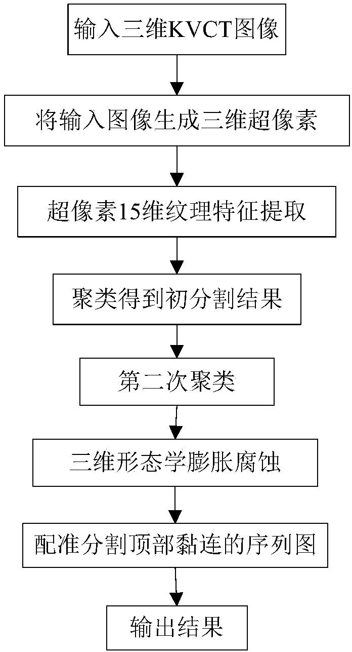
[0032] Reference figure 1 , The implementation steps of the present invention are as follows:

[0033] Step 1. Enter the KVCT three-dimensional image D of the prostate to be segmented, such as figure 2 Shown is a typical cross-section selected from the KVCT three-dimensional drawing.

[0034] Step 2. Use the SLIC super pixel generation algorithm to generate a three-dimensional matrix S composed of three-dimensional super pixels on the three-dimensional KVCT image D of the prostate.

[0035] 2a) Initialize the three-dimensional matrix S to have the same size as D;

[0036] 2b) The initial size of each super pixel is 5×5×5, with a total of 125 pixels. If there are N pixels in D, set the number of super pixels to be divided into n=N / 125;

[0037] 2c) Perform the SLIC operation on D to get the final S. The label of the corresponding superpixel is stored in S, and the pixels contained in each superpixel are marked with the same number;

[0038] Step 3. Calculate the 15-dimensional texture f...


PUM


Abstract

The present invention discloses a prostate KVCT image prostate region 3D segmentation method, which mainly aims to solve the problem of failing to perform prostate segmentation on a KVCT image. The implementation steps of the method are as follows: 1) inputting a to-be-segmented prostate KVCT three-dimensional image, and generating super pixels based on the three-dimensional image; 2) calculating a 15-dimensional texture feature of the super pixels; 3) performing primary clustering on the super pixels to obtain an initial segmentation result; 4) performing secondary clustering on the initial segmentation result to obtain two clustering results; 5) performing three-dimensional morphological processing on the two clustering results to obtain an adhesion-removing result; and 6) segmenting an image with severe adhesion at the top of a sequence graph by using registration, and then substituting an obtained part of the sequence graph by registration segmentation for the corresponding sequence graph in the adhesion-removing graph, so as to obtain a final segmentation result.

Description

Technical field [0001] The invention belongs to the field of image processing technology, relates to a medical CT image processing method, and can be used for segmentation of prostate KVCT images. Background technique [0002] The prostate is the largest accessory sex gland in men and is also one of the secretory glands outside the human body. It is located between the bladder and the genital diaphragm. Prostate cancer is a malignant tumor arising from the prostate. Gene mutations in the cells lead to uncontrolled proliferation and become cancer. In addition to expanding in size or invading neighboring organs, malignant cells may also metastasize to other parts of the body, especially bones and lymph nodes. Prostate cancer may cause symptoms such as pain, difficulty urinating, and erectile dysfunction. In Western countries, prostate cancer is the second most common cancer in men, so the number of people killed is second only to lung cancer. The extraction of prostate lesions i...

Claims


Application Information

Patent Timeline
Patent Type & Authority Patents(China)
IPC IPC(8): G06T7/11G06T7/187
CPCG06T7/0012G06T2207/10081G06T2207/30081
Inventor 姚瑶杨光缑水平
Owner HANGZHOU VOCATIONAL & TECHN COLLEGE
Why Eureka
Eureka Blog
Learn More