A directional differentiation induction method of human embryonic stem cells to obtain corneal endothelial cells

A technology of human embryonic stem cells and induced differentiation, which is applied in the field of directional induction and differentiation of human embryonic stem cells into corneal endothelial cells.

Active Publication Date: 2016-11-30
GENERAL HOSPITAL OF PLA
View PDF3 Cites 14 Cited by
  • Summary
  • Abstract
  • Description
  • Claims
  • Application Information

AI Technical Summary

Problems solved by technology

Due to the limited proliferation ability of corneal endothelial cells, it is difficult to culture and passage corneal endothelial c

Method used


Image

  • A directional differentiation induction method of human embryonic stem cells to obtain corneal endothelial cells
  • A directional differentiation induction method of human embryonic stem cells to obtain corneal endothelial cells
  • A directional differentiation induction method of human embryonic stem cells to obtain corneal endothelial cells

Examples


Embodiment Construction

[0043] The following examples are used to illustrate the present invention, but are not intended to limit the scope of the present invention. Unless otherwise specified, the technical means used in the examples are conventional means well known to those skilled in the art, and the raw materials used are all commercially available products.

[0044] Embodiment The method for directional induction and differentiation of human embryonic stem cells into corneal endothelial cells

[0045] The method comprises the steps of:

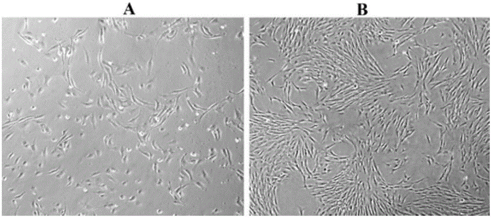
[0046] S1. Human embryonic stem cells differentiated into human neural crest stem cells

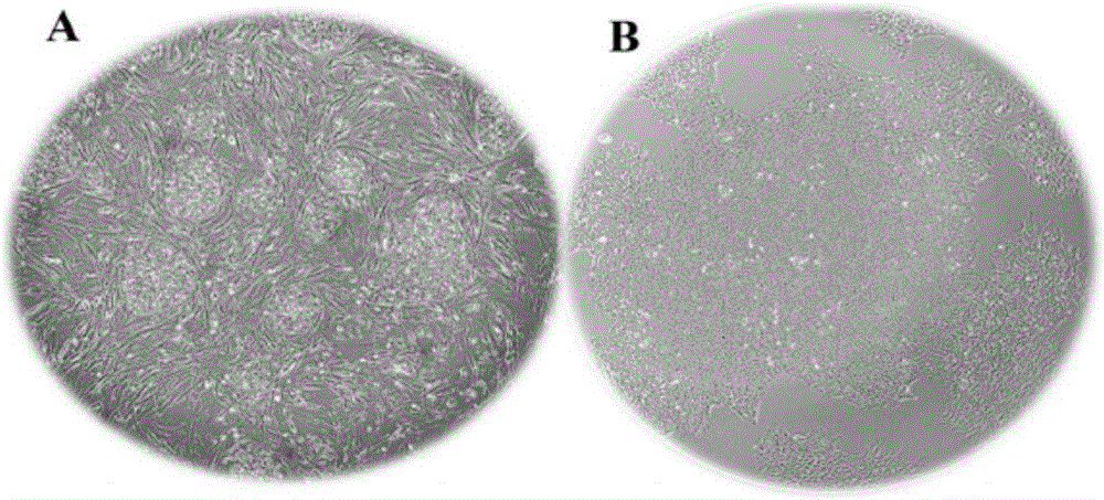
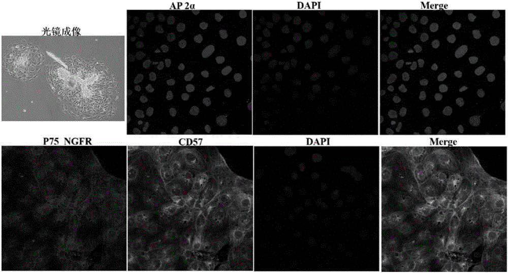
[0047] S2. Human neural crest stem cells were induced to differentiate into human corneal endothelial cells

[0048] Preferably, after step S1, the method further includes the step of sorting induced differentiation cells by flow cytometry, and collecting cells showing positive expression of neural crest stem cell markers.

[0049] Step S1 is as follows: 37 ° C, 5% CO ...


PUM


Abstract

A directional differentiation induction method of human embryonic stem cells to obtain corneal endothelial cells is provided. The method includes inducing the human embryonic stem cells to differentiate into human neural crest stem cells, inducing the human neural crest stem cells to differentiate into the human corneal endothelial cells, and other steps. The method adopts a primary human corneal stroma cell culture supernatant fluid, a human corneal endothelial cell culture supernatant fluid and a human lens cell culture supernatant fluid as a conditioned medium. Through adding retinoic acid and a plurality of recombinant proteins into the medium, and combining three-dimensional and two-dimensional culture methods, a corneal endothelial cell development process is simulated to induce the human embryonic stem cells to directionally differentiate into the human corneal endothelial cells, the corneal endothelial cells morphological structures of which are similar to those of normal human corneal endothelial cells can be obtained, and in-vitro subculture results show that proliferation of the obtained human corneal endothelial cells is consistent with proliferation of the normal human corneal endothelial cells.

Description

technical field [0001] The invention relates to the field of cell biology, in particular to a method for directional induction and differentiation of human embryonic stem cells into corneal endothelial cells. Background technique [0002] The cornea is located at the front of the eyeball and is divided into five layers from front to back, which are epithelial cell layer, Bowman's layer, stroma layer, Bowman's layer and endothelial cell layer. Corneal disease is one of the common blinding eye diseases in my country, which seriously affects the visual quality and quality of life of patients. At present, there are two main treatment methods for various corneal diseases that affect the function of the corneal endothelium, Descemetmembrane endothelial keratoplasty (DMEK) and Descemet's stripping automated endothelial keratoplasty (DSAEK). Compared with traditional full-thickness keratoplasty (penetratingkeratoplasty, PK), endothelial keratoplasty has a better prognosis and mild ...

Claims


Application Information

Patent Timeline
IPC IPC(8): C12N5/079C12N5/0797
CPCC12N5/0621C12N5/0623C12N2500/38C12N2501/11C12N2501/115
Inventor 陈小鸟吴玲玲王丽强黄一飞
Owner GENERAL HOSPITAL OF PLA
Why Eureka
Try Eureka