

HIV-1 protease detection method based on solid state nanopore
A HIV-1 and nanopore technology, applied in the field of precision medical research, can solve the problems of heavy workload and time-consuming
- Summary
- Abstract
- Description
- Claims
- Application Information
AI Technical Summary
Benefits of technology
Problems solved by technology
Method used
Image

Examples
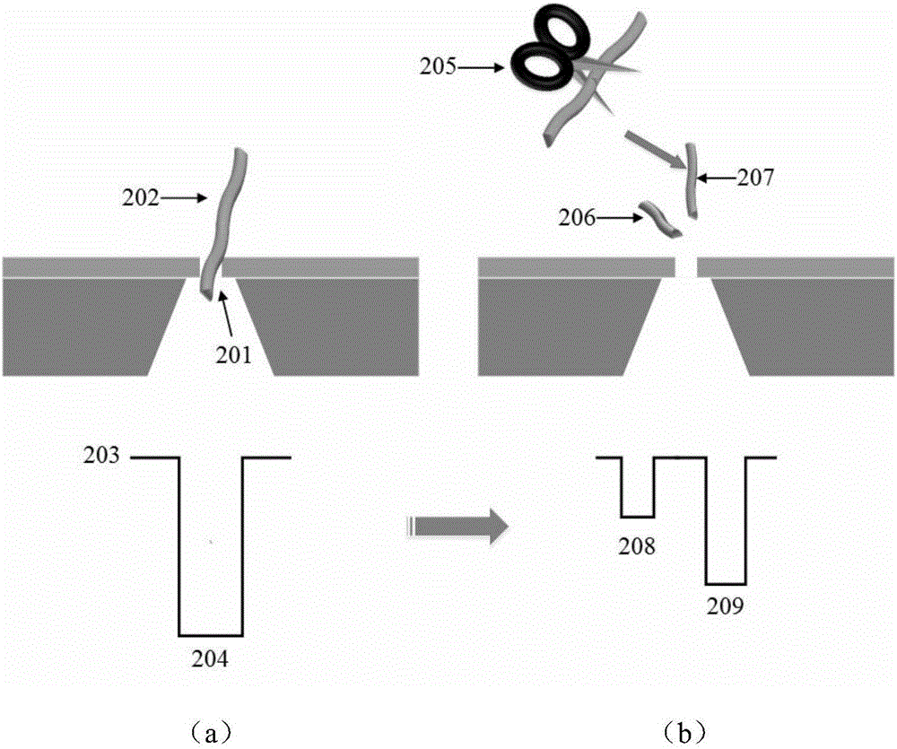
Embodiment 1
[0025] 1. Provide solid-state nanopore detection system
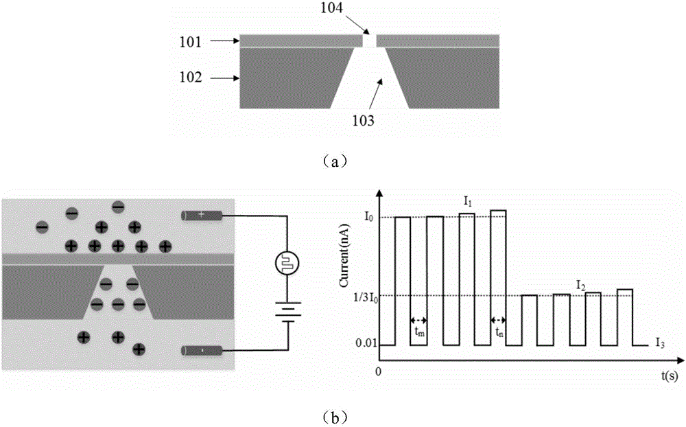
[0026] The solid-state nanopore detection system of the present invention includes a solid-state nanopore sensor, a salt solution chamber and a current monitoring device. The solid-state nanopore sensor includes: a substrate 102 and an electrically insulating film 101 compounded on the substrate 102. The substrate 102 is provided with a backside window 103. The electrically insulating film 101 and the top of the backside window 103 are etched with nanometers. The hole 104, the back window 103 and the nano hole 104 are located inside the salt solution chamber and communicate with the upper chamber and the lower chamber; the current monitoring device includes an electrically connected power supply, a current meter, an electrode I, an electrode II, the electrode I , Electrode II is located in the upper chamber and the lower chamber respectively.
[0027] The structure diagram of the solid-state nanopore sensor is as follows fi...
PUM
| Property | Measurement | Unit |
|---|---|---|
| Thickness | aaaaa | aaaaa |
| Diameter | aaaaa | aaaaa |
| Thickness | aaaaa | aaaaa |
Abstract
Description
Claims
Application Information