Method for efficiently amplifying subgroup J avian leukosis virus (ALV)

A technology of avian leukosis virus and subgroups, applied in the field of immunology, can solve the problems of time-consuming, etc., and achieve the effect of improving sensitivity and shortening the separation and detection time

Active Publication Date: 2018-01-19
YANGZHOU UNIV
View PDF1 Cites 4 Cited by
  • Summary
  • Abstract
  • Description
  • Claims
  • Application Information

AI Technical Summary

Benefits of technology

This patented technology allows birds (chick) that are infected by Leucovirus disease or other diseases like malaria have been isolated from their bloodstreams before they develop symptoms such as encephalitis. By detecting this new type of virus, researchers may better diagnose these conditions more accurately than previous techniques which took several months to study them.

Problems solved by technology

This patented describes how an organism called Chikungunya Virus affects birds through various ways such as transmitting disease symptoms like abnormal growth rates caused by certain genotypes of CHIKEN VILLARS virus. However, despite being isolated during testing, some individuals may still develop latently fatal necrosis syndrome due to their own gene mutations. There currently does not exist any effective way to detect and treat Alvirus infections effectively without relying solely upon laborious methods involving culturing live donor organs collected after several months' suspension period.

Method used


Image

  • Method for efficiently amplifying subgroup J avian leukosis virus (ALV)
  • Method for efficiently amplifying subgroup J avian leukosis virus (ALV)

Examples


Embodiment Construction

[0021] The present invention will be further described below in conjunction with specific examples.

[0022] 1) Cell culture

[0023] Chicken hepatocytes were cultured in DMEM medium containing 10% fetal bovine serum, and DF-1 cells were cultured in DMEM medium containing 5% fetal bovine serum. After the cells covered the monolayer, they were washed once with PBS. Digest with 0.25% trypsin and pass to 6-well or 96-well cell culture plate.

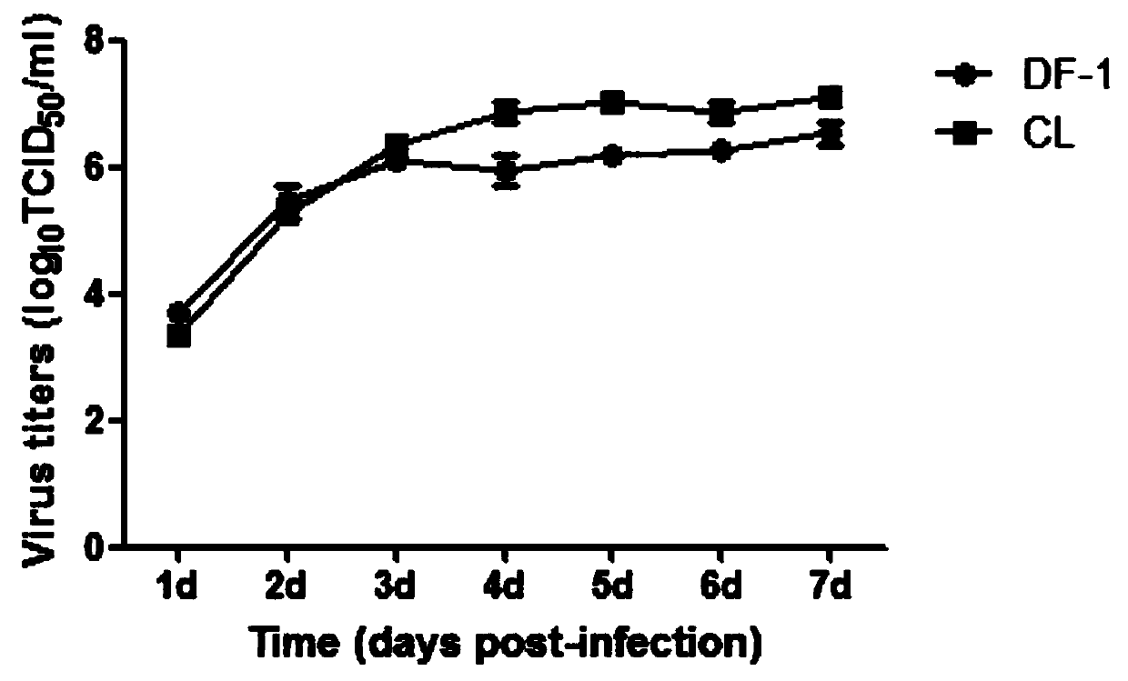
[0024] 2) Efficient amplification of subgroup J avian leukemia virus in chicken hepatocytes

[0025] Chicken hepatocytes and DF-1 cells were inoculated into a 6-well plate for culture. When the confluence was about 70%, the J subgroup avian leukosis virus GY03 strain was inoculated into the two kinds of cells, and the MOI was 0.001. Repeatedly, cultivated with 2ml medium containing 2% fetal bovine serum for 7 days, collected 200 μl supernatant every day and stored it in a -80°C refrigerator for later use, and supplemented 200 μl medium co...


PUM


Abstract

The invention relates to a method for efficiently amplifying subgroup J avian leukosis virus (ALV). According to the method, chicken hepatocytes can be used for efficiently amplifying the subgroup J ALV, the characteristic of high titer virus can be produced earlier by using the chicken hepatocytes, the separation and detection time of the subgroup J ALV can be shortened, the pathogen can be detected earlier, and the sensitivity of a subgroup J ALV detection method is improved. When subgroup J ALV infection MOI is 0.001 hour, the effect of enzyme-linked immunosorbent assay (ELISA) on the chickhepatocytes infected for 3 days is comparable to the effect of ELISA on DF-1 cells infected for 7 days. The efficient amplification method established by the invention accelerates the purification process of the subgroup J ALV, thus shortening the time and reducing cost; the method has very good application value in the purification detection of the subgroup J ALV.

Description


Claims


Application Information

Patent Timeline
Owner YANGZHOU UNIV
Why Eureka
Try Eureka