Image-guide-type fundus camera

An image-guided and camera-based technology, applied in ophthalmoscopes, eye testing equipment, medical science, etc., can solve the problem that doctors cannot understand the condition of the user's fundus

Pending Publication Date: 2018-08-21
合肥奥比斯科技有限公司
View PDF0 Cites 0 Cited by
  • Summary
  • Abstract
  • Description
  • Claims
  • Application Information

AI Technical Summary

Problems solved by technology

[0005] At present, the fundus cameras on the market use light beams to illuminate the fundus and output the fundus image. This method can only capture fundus images in one direction, so that doctors cannot fully understand the fundus of the user.

Method used


Image

  • Image-guide-type fundus camera

Examples


Embodiment Construction

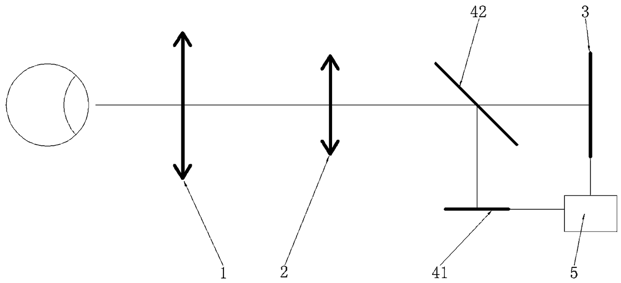
[0014] Such as figure 1 As shown, in this embodiment, the present invention includes a retinal objective lens 1, an imaging objective lens 2 and an image sensor 3 arranged sequentially on the same axis, and the fundus camera also includes an image generator, and the image generator includes a display 41 and a beam splitter 42, the beam splitter 42 is arranged between the imaging objective lens 2 and the image sensor 3, the display 41 cooperates with the beam splitter 42, and the image beam emitted by the display 41 passes through the After being reflected by the spectroscope 42, the fundus is illuminated through the imaging objective lens 2 and the retinal objective lens 1, and an image is formed on the imaging objective lens 2 at the same time, and the size and position of the image generated by the display 41 are adjusted to guide human eyes to observe direction and its focus. The image sensor 3 is a charge-coupled device image sensor 3 . The fundus camera further includes...


PUM


Abstract

The invention provides an image-guide-type fundus camera for achieving focusing and human eye positioning through the human eyes. The camera comprises an omentum objective lens (1), an imaging objective lens (2) and an image sensor (3) which are arranged on the same axis in sequence, the fundus camera further comprises an image generator, and the image generator comprises a displayer (41) and a spectroscope (42); the spectroscope (42) is arranged between the imaging objective lens (2) and the image sensor (3), the displayer (41) is matched with the spectroscope (42), and an image light beam sent by the displayer (41) is reflected by the spectroscope (42) and then illuminates the fundi through the imaging objective lens (2) and the omentum objective lens (1); meanwhile, an image is formed on the imaging objective lens (2), the size and position of the image generated by the displayer (41) are adjusted, and then the observation direction and focus of the human eye are guided. The image-guide-type fundus camera is applied to the technical field of fundus cameras.

Description

technical field [0001] The invention relates to a fundus camera, in particular to an image-guided fundus camera. Background technique [0002] The cornea, iris, lens, etc. in front of the eyeball are defined as the anterior segment by the ophthalmology field, and the vitreous body, retina, choroid, etc. in the back are defined as the posterior segment. The bottom of the posterior segment of the eye is called the fundus. The fundus is composed of the retina, fundus blood vessels, optic nerve head, optic nerve fibers, macula on the retina, and choroid behind the retina. The lesions in these parts are collectively called fundus diseases. [0003] The fundus is the only place where arteries, veins and capillaries can be directly and concentratedly observed with the naked eye. These blood vessels can reflect the dynamics and health status of the blood circulation throughout the body. Therefore, fundus examination is not only an important method to check eye vitreous, retina, ch...

Claims


Application Information

Patent Timeline
Patent Type & Authority Applications(China)
IPC IPC(8): A61B3/12A61B3/14
CPCA61B3/12A61B3/0041A61B3/14
Inventor 闫佩正陆宇贠新乐孙明斋
Owner 合肥奥比斯科技有限公司
Why Eureka
Try Eureka