
Identifying candidate cells using image analysis

A cell and candidate target technology, which can be used in image analysis, image enhancement, analysis of materials, etc., and can solve the problems of difficulty in accurate detection and counting of CTCs

Active Publication Date: 2018-10-23
CELLMAX LTD
View PDF13 Cites 11 Cited by
  • Summary
  • Abstract
  • Description
  • Claims
  • Application Information

AI Technical Summary

Problems solved by technology

Therefore, CTCs are difficult to detect and count accurately

Method used


Image

  • Identifying candidate cells using image analysis
  • Identifying candidate cells using image analysis
  • Identifying candidate cells using image analysis

Examples


Embodiment Construction

[0035] The technical solutions of the present invention will be clearly and completely described below in conjunction with the accompanying drawings. Apparently, the described embodiments are part of the embodiments of the present invention, not all of them. Based on the embodiments of the present invention, all other embodiments obtained by persons of ordinary skill in the art without making creative efforts belong to the protection scope of the present invention.

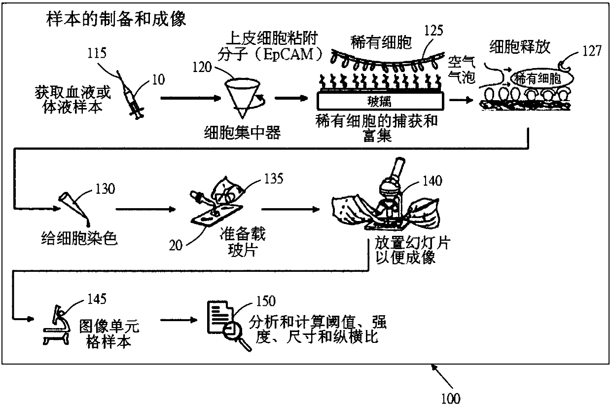
[0036]Solid tumor sampling is a routine procedure in cancer diagnosis. Today, next-generation sequencing technologies enable sensitive, rapid, and low-cost detection and analysis of tumor DNA in cancer cells or their constituent DNA that has progressed beyond its tissue of origin into fluid components between cells (e.g., Interstitial fluid, lymph fluid, blood, saliva, cerebrospinal fluid, synovial fluid, urine, feces and other secretions). Fragments of cancer cells harvested from primary tumors can serve as mark...


PUM

PropertyMeasurementUnit
sizeaaaaaaaaaa
pore sizeaaaaaaaaaa

Abstract

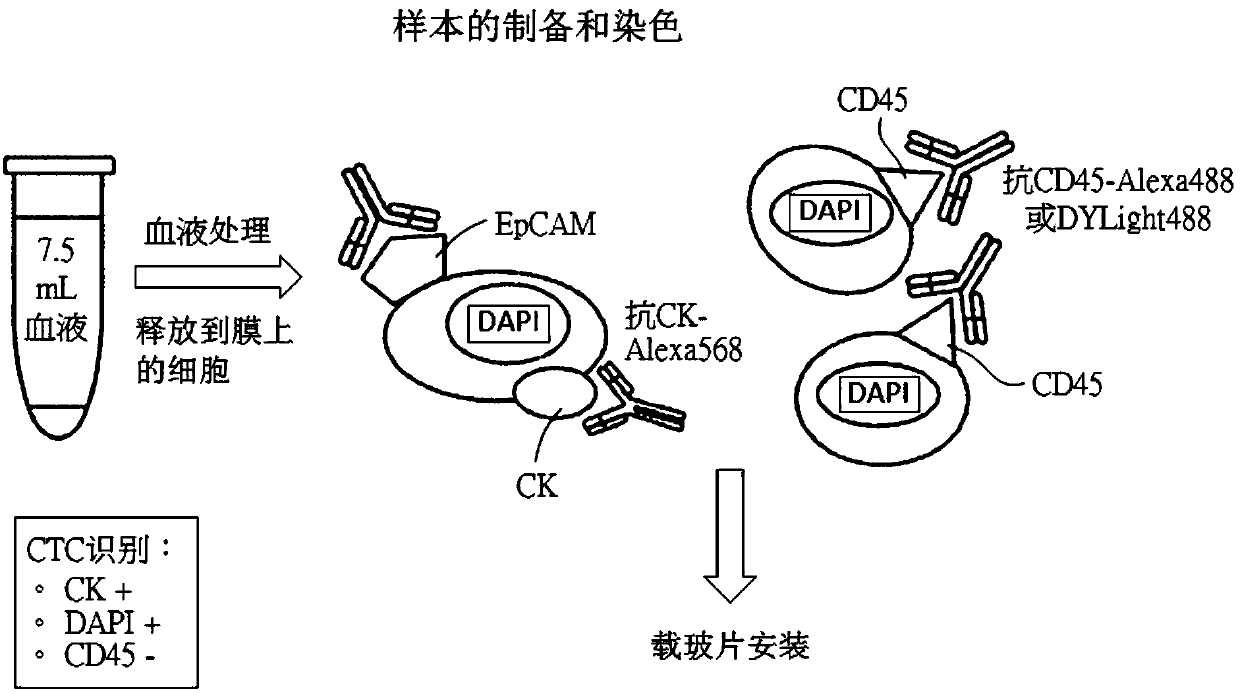
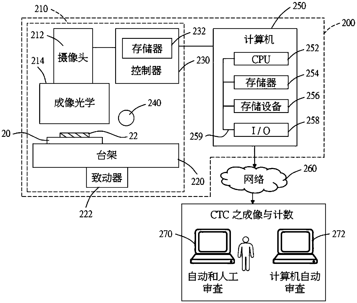
A method for identifying and counting candidate target cells within a biological fluid specimen is described. The method includes obtaining a biological fluid specimen, preparing the biological fluidspecimen by staining cell features in the biological fluid specimen, capturing a digital image having a plurality of color channels of the biological fluid specimen, and applying image analysis to thedigital image. A computer program product for identifying candidate target cells within a biological fluid specimen is also described. The computer program comprises instructions to cause a processorto carry out the image analysis. In specimen areas possibly including interesting candidate cells, the areas can be positioned automatically, the areas of this type can be labeled for further evaluation, and the number of specimen sample areas requiring checking by an operator is obviously reduced. The method is used for CTC detection and counting automatic imaging, can facilitate prediction of disease development and the total survival time during a treatment period, and can achieve continuous monitoring on prognosis of patients, thus providing a smarter patient nursing choice.

Description

technical field [0001] The invention relates to identifying candidate cells (eg, circulating tumor cells) in images of a sample. Background technique [0002] Circulating tumor cells (CTCs) are cancer cells that have shed from the primary tumor and entered the vasculature or lymphatic system, usually of epithelial origin. When CTCs invade the circulation, such malignant cells enter other organs. After breaking off from the solid block, CTCs may lodge on the vessel wall and infiltrate the surrounding tissue. Angiogenesis promotes the development of new tumors at sites distant from the original mass. Thus, CTCs represent the seeds from which other tumors (metastases) grow. [0003] It is understood that the number of CTCs in peripheral blood is associated with shortened progression-free survival and overall survival in patients with metastatic disease, including breast, colorectal and prostate cancers. CTC detection and enumeration from blood or other bodily fluid samples ...

Claims


Application Information

Patent Timeline
Patent Type & Authority Applications(China)
IPC IPC(8): G01N1/30G01N15/10G06T7/90G06T7/00
CPCG06T7/0012G06T2207/30096G01N1/31G01N15/1475G01N33/5094G01N33/57484G01N33/582G01N2333/4742G01N2333/70589G06T2207/30242G06T2207/10056G06T2207/10064C12Q1/6886C12Q1/6883G01N15/1468G01N2015/1472G06T2207/30101
Inventor 谢煌彬赖志铭梅蕊邵宏仁王小明吴仁家
Owner CELLMAX LTD
Learn More