Integral enhancement method for digital subtraction angiography image

A technique of vascular imaging and digital subtraction, applied in image enhancement, image analysis, image data processing and other directions, can solve problems such as increasing patient pain, high-frequency loss of images, and reducing image contrast, reducing injection volume and collecting images. time, suppress random noise, enhance the effect of display function

Active Publication Date: 2018-11-06
SOUTHEAST UNIV
View PDF7 Cites 4 Cited by
  • Summary
  • Abstract
  • Description
  • Claims
  • Application Information

AI Technical Summary

Problems solved by technology

However, due to the limited frame rate of DSA images and the injection concentration of contrast agent should not be too high, the contrast of DSA images is low, which makes clinical diagnosis difficult
At the same time, in order to reduce breathing artifacts, the patient is usually in a state of respiratory arrest during the shooting process, which increases the pain of the patient
[0003] Currently commonly used image denoising and super-resolution technologies mainly include compressed sensing technology, total variation technology, and wavelet decomposition and reconstruction technology. Real-time requirements; wavelet decomposition and reconstruction technology will cause the loss of high-frequency parts of the image, further reducing the contrast of the image
[0004] In addition, since blood flow is a periodic dynamic process, a single picture cannot contain all the information doctors need

Method used


Image

  • Integral enhancement method for digital subtraction angiography image
  • Integral enhancement method for digital subtraction angiography image
  • Integral enhancement method for digital subtraction angiography image

Examples


Embodiment Construction

[0020] The present invention will be described in further detail below in conjunction with the accompanying drawings and embodiments. In this embodiment, the imaging equipment used is: PHILIPS AlluraXper FD20; the contrast agent is: Omnipaque (iodine content specification: 350mg / ml, dosage 100ml), which is a product of (General Electric Pharmaceutical (Shanghai) Co., Ltd.). The method of contrast medium angiography was as follows: a catheter was punctured through the femoral artery, and a high-pressure syringe (Medrad Mark V Provis) was used to inject contrast medium into the aorta through a 5F gold-marked pigtail catheter. The filming speed was 6 frames / s, and the injection pressure was 600 psi (4136.85 kPa). In this embodiment, the software used in the algorithm is Matlab2010.

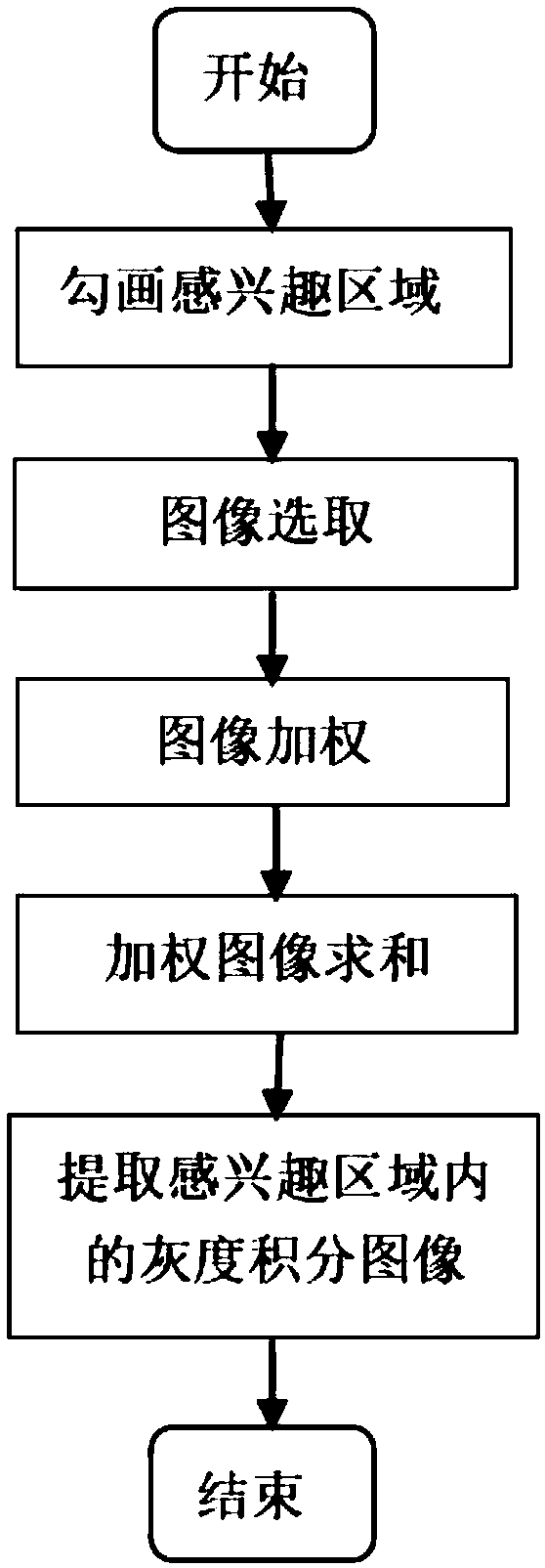
[0021] Such as figure 1 , the integral enhancement method of the digital subtraction angiography image of the present invention comprises the following steps:

[0022] (1) Delineate the region of ...


PUM


Abstract

The invention discloses an integral enhancement method for a digital subtraction angiography image. The method includes the following steps that: (1) a region of interest is delineated in a DSA (digital subtraction angiography) image; (2) a plurality of images in a first cardiac cycle which are obtained after a contrast agent enters the region of interest of a blood vessel, and the images are de-noised; (3) the weighted images of the plurality of de-noised images are obtained based on shooting time intervals between the plurality of images; and (4) the weighted images are summed, so that a grayscale integrated image within the cardiac cycle is obtained. With the method of the invention adopted, the image enhancement of the digital subtraction angiography image can be realized, and therefore, doctors can be facilitated to locate and distinguish some characteristic positions in the blood vessel, and the reduction of the injection dose of the contrast agent for a patient can be facilitated, and image acquisition time can be shortened.

Description

technical field [0001] The invention relates to an image enhancement and denoising method, in particular to an integral enhancement method for digital subtraction angiography images. technical background [0002] Digital subtraction angiography (DSA) method is a commonly used auxiliary diagnosis method, which has good real-time performance and is convenient for real-time monitoring of patients during operation. However, due to the limited frame rate of DSA images and the injection concentration of contrast agent should not be too high, the contrast of DSA images is low, which makes clinical diagnosis difficult. At the same time, in order to reduce breathing artifacts, the patient is usually in a breathing stop state during the shooting process, which increases the pain of the patient. [0003] Currently commonly used image denoising and super-resolution technologies mainly include compressed sensing technology, total variation technology, and wavelet decomposition and recon...

Claims


Application Information

Patent Timeline
Patent Type & Authority Applications(China)
IPC IPC(8): G06T5/00G06T5/50
CPCG06T5/002G06T5/007G06T5/50G06T2207/10116G06T2207/30101
Inventor 黄朔万遂人孙钰周平朱斌程乐张冰
Owner SOUTHEAST UNIV
Why Eureka
Try Eureka