
Pulmonary ultrasound image analysis method and apparatus, electronic device and readable storage medium

A technology of ultrasound images and analysis methods, applied in the field of data processing, which can solve problems such as unproposed solutions and the impact of ultrasound examination results

Active Publication Date: 2018-11-20
ZHEJIANG UNIV
View PDF8 Cites 19 Cited by
  • Summary
  • Abstract
  • Description
  • Claims
  • Application Information

AI Technical Summary

Problems solved by technology

To complete the examination of a target organ, the operator needs to perform multiple scans from different parts and angles, which greatly affects the results of ultrasound examination
[0004] For the above problems, no effective solution has been proposed

Method used

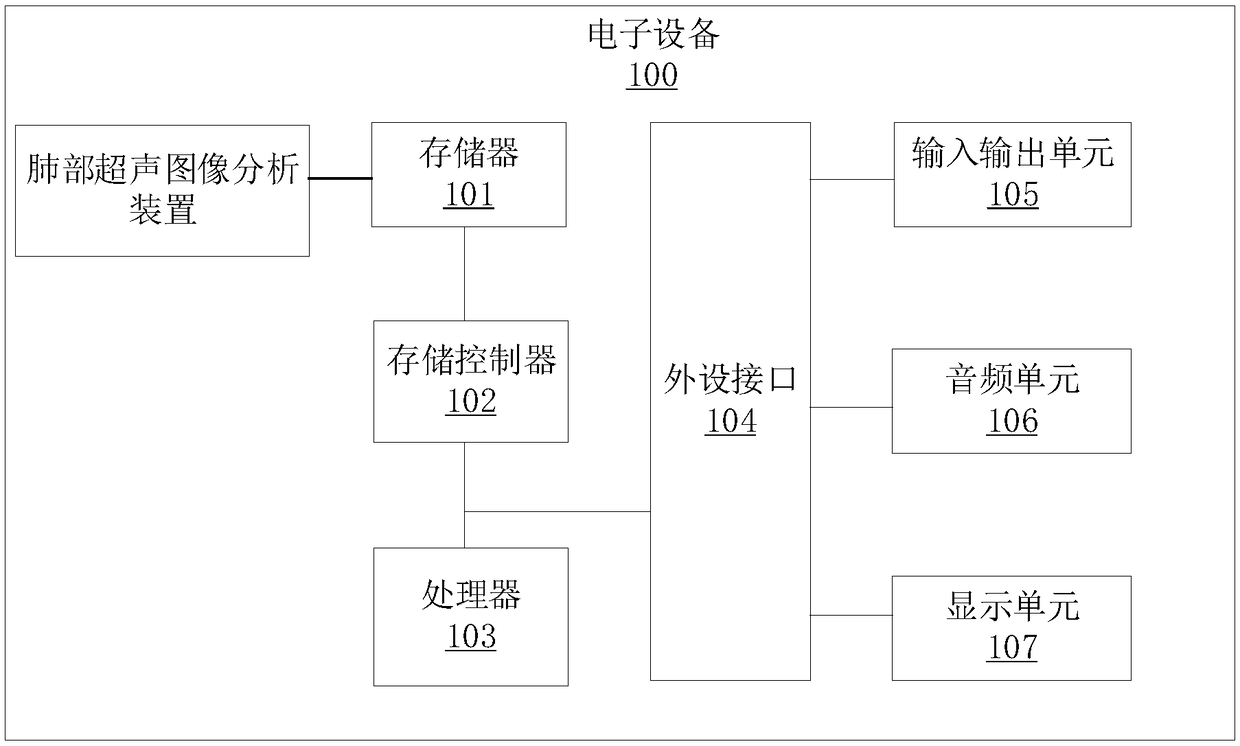
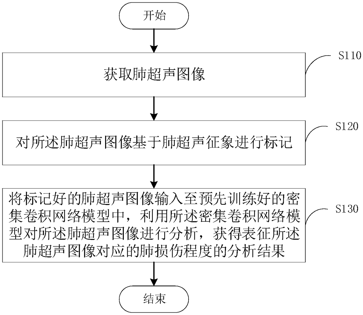
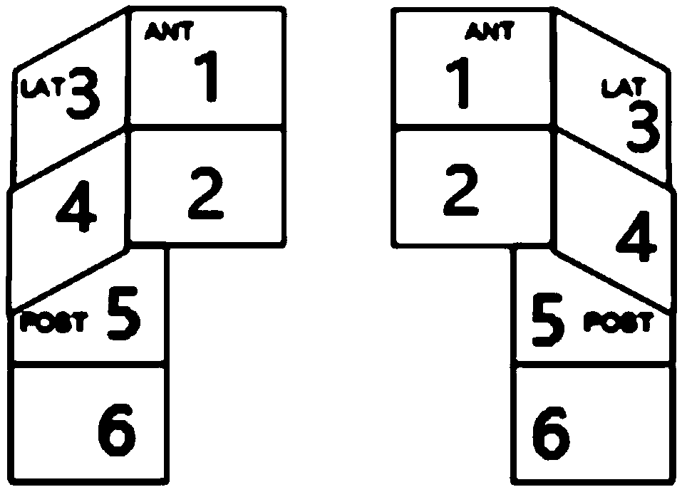

Image

  • Pulmonary ultrasound image analysis method and apparatus, electronic device and readable storage medium
  • Pulmonary ultrasound image analysis method and apparatus, electronic device and readable storage medium
  • Pulmonary ultrasound image analysis method and apparatus, electronic device and readable storage medium

Examples


Embodiment Construction

[0043] The following will clearly and completely describe the technical solutions in the embodiments of the present invention with reference to the accompanying drawings in the embodiments of the present invention. Obviously, the described embodiments are only some, not all, embodiments of the present invention. The components of the embodiments of the invention generally described and illustrated in the figures herein may be arranged and designed in a variety of different configurations. Accordingly, the following detailed description of the embodiments of the invention provided in the accompanying drawings is not intended to limit the scope of the claimed invention, but merely represents selected embodiments of the invention. Based on the embodiments of the present invention, all other embodiments obtained by those skilled in the art without making creative efforts belong to the protection scope of the present invention.

[0044] It should be noted that like numerals and let...


PUM


Abstract

The embodiment of the invention provides a pulmonary ultrasound image analysis method and apparatus, an electronic device and a readable storage medium. The method comprises the following steps: obtaining a pulmonary ultrasound image, then marking the pulmonary ultrasound image based on a pulmonary ultrasound sign, then inputting the marked pulmonary ultrasound image into a pre-trained dense convolutional network model, and analyzing the pulmonary ultrasound image by using the dense convolutional network model to obtain an analysis result of a lung injury degree corresponding to the pulmonaryultrasound image. The obtained pulmonary ultrasound image is analyzed by using the pre-trained dense convolutional network model, the obtained analysis result can reflect the lung injury degree corresponding to the pulmonary ultrasound image, therefore, the purpose of characterizing the lug health condition according to the analysis result is achieved, and the technical problem that the ultrasoundimage cannot be analyzed and processed to study the health state of a patient based on the analysis result in the prior art is solved.

Description

technical field [0001] The present invention relates to the field of data processing, in particular to a lung ultrasound image analysis method, device, electronic equipment and readable storage medium. Background technique [0002] The application of ultrasound technology skills in clinical departments has increasingly become a basic skill that clinicians care about. In many clinical departments, especially in perioperative and critical care related departments, ultrasound examination has gradually become a routine skill for clinicians. Lung ultrasonography is the core of ultrasonography in emergency and critical care. Unlike other traditional ultrasonography, lung ultrasonography does not reflect direct images of lung tissue in most cases, but a series of artifacts. These artifacts are defined by the display characteristics of lung ultrasound images. [0003] But ultrasonography also has its downsides. The results of ultrasonography are largely influenced by the skill of...

Claims


Application Information

Patent Timeline
IPC IPC(8): G06T7/00
CPCG06T7/0012G06T2207/10132G06T2207/20081G06T2207/30061
Inventor 张茂吴春双
Owner ZHEJIANG UNIV
Learn More