Thyroid gland regional automatic segmentation method based on CT image

A technology for automatic segmentation of CT images, applied in the field of medical imaging, can solve problems such as low efficiency and inability to meet automatic diagnosis, and achieve the effect of meeting technical requirements

Active Publication Date: 2019-02-19
ZHEJIANG MEDICAL COLLEGE
View PDF7 Cites 4 Cited by
  • Summary
  • Abstract
  • Description
  • Claims
  • Application Information

AI Technical Summary

Problems solved by technology

Although this method extracts the most accurate boundaries, it is ex

Method used


Image

  • Thyroid gland regional automatic segmentation method based on CT image
  • Thyroid gland regional automatic segmentation method based on CT image
  • Thyroid gland regional automatic segmentation method based on CT image

Examples


Embodiment Construction

[0025] The present invention will be further described in detail below in conjunction with the drawings, but it should not be understood that the scope of the above-mentioned subject of the present invention is limited to the above-mentioned embodiments.

[0026] Such as Image 6 As shown, an automatic segmentation method of thyroid region based on CT images includes the following steps:

[0027] 1. Obtain images and registration of transverse position before and after thyroid enhancement:

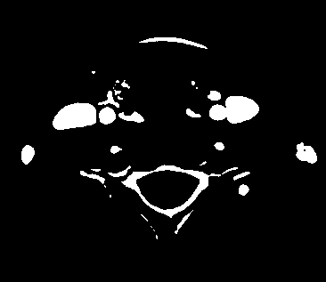
[0028] The pre-enhanced image refers to the plain scan image, that is, the transverse image obtained by scanning the vein without iodine injection, such as figure 1 As shown, neck blood vessels, thyroid and other tissues do not contain iodine contrast agent, blood vessels, thyroid and soft tissues do not contain iodine contrast agent, normal thyroid, the tissue density is slightly higher than the surrounding soft tissue, there are obvious boundaries;

[0029] The enhanced image refers to the cross-...


PUM


Abstract

The invention belongs to the field of medical imaging, in particular to a CT image segmentation method. The method comprises the steps that 1, cross section position images before and after thyroid gland enhancement and registering is conducted; 2, a thyroid gland tissue template extracted; 3, thyroid tissues are segmented. The method has the advantages of high speed, accurate and the like, and the technical demands of thyroid gland CT image automatic diagnosis can be met.

Description

Technical field [0001] The invention belongs to the field of medical imaging, and specifically relates to a CT image segmentation method. Background technique [0002] CT examination is one of the important methods for the diagnosis of thyroid diseases. Imaging surgeons can use CT scans to find abnormal densities in the thyroid gland, such as high-density calcifications, low-density necrotic areas, or cystic lesions, to determine the nature of thyroid lesions. At the same time, in order to obtain more internal information of the thyroid and better distinguish diseased tissues with a density similar to the normal thyroid, the method of thyroid enhancement is often used, that is, the patient needs to undergo multiple CT scans after intravenous injection of iodine contrast agent. Due to the different metabolic functions of iodine contrast agent between diseased tissue and normal thyroid tissue, the concentration of polymer iodine compound in normal thyroid and diseased tissue will ...

Claims


Application Information

Patent Timeline
IPC IPC(8): A61B6/00
CPCA61B6/481A61B6/5241
Inventor 彭文献彭天舟王上东田慧彭恰恰陈益红
Owner ZHEJIANG MEDICAL COLLEGE
Why Eureka
Try Eureka