Visual pharyngoscope device

A technology of laryngoscope and tongue depressor, applied in laryngoscope, bronchoscope, medical science, etc., to solve difficult airway intubation problems, eliminate camera blind spots, and facilitate gastric tube or transesophageal ultrasound probe

Inactive Publication Date: 2019-06-11
蔡丽丽
View PDF7 Cites 0 Cited by
  • Summary
  • Abstract
  • Description
  • Claims
  • Application Information

AI Technical Summary

Problems solved by technology

[0003] Aiming at the problems existing in the above-mentioned prior art, the present invention provides a visual laryngoscope device, which can combine the advantages of traditional lenses and visual systems to better perform endotracheal intubation, and the attachment position and camera angle can be adjusted for better Eliminate camera blind spots and better solve difficult airway intubation problems

Method used


Image

  • Visual pharyngoscope device
  • Visual pharyngoscope device

Examples


Embodiment Construction

[0011] The present invention will be further described below in conjunction with the accompanying drawings.

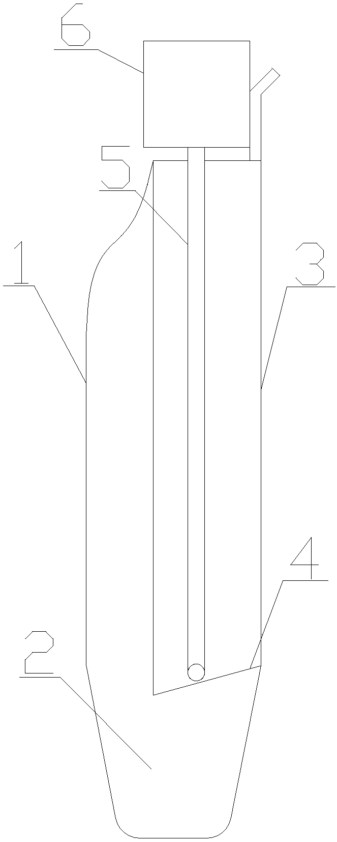
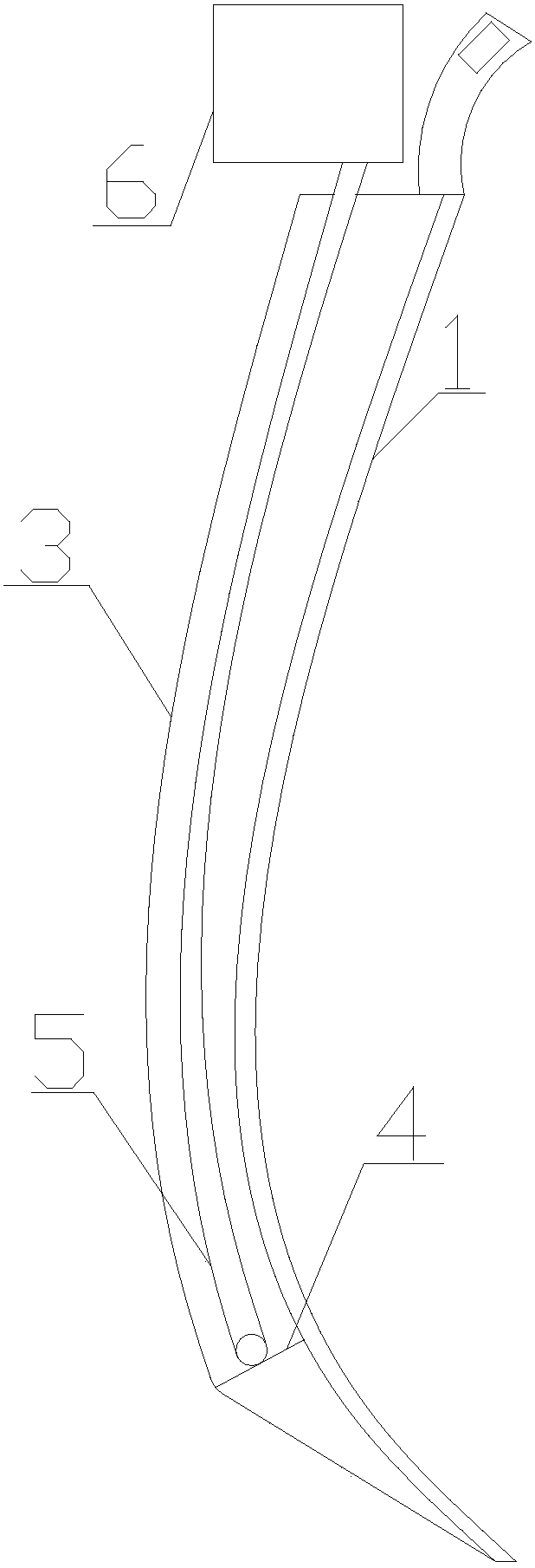
[0012] Such as figure 1 with figure 2 As shown, the video laryngoscope device includes a tongue depressor 1, a top end 2, a flange 3, a top surface 4, a transmission tube 5 and a joint 6, the spatula 1 and the flange 3 are injection molded at one time, and the tongue depressor 1 and flange 3 have no edges and corners and no burrs. The tongue depressor 1 is on the left side of the flange 3, and the top 2 is a transparent panel. The rectangular flange 3 protrudes from the spatula 1 by one to two centimeters. A strip-shaped transmission tube 5 is attached to the middle position, and an adhesive layer is pasted on one side of the transmission tube 5, which can be attached to the lens of a traditional invisible anesthesia laryngoscope, or placed in a speculum similar to a tracheal tube and a bronchoscope One end of the strip-shaped transmission tube 5 is a camera, and th...


PUM


Abstract

The invention relates to a visual pharyngoscope device, and belongs to the field of medical apparatuses. The visual pharyngoscope device comprises a tongue depressor (1), a top end (2), an outstandingflange (3), a top surface (4), a transmission pipe (5) and a combining part (6), wherein the tongue depressor and the outstanding flange are integrally injected and shaped, the periphery of the tongue depressor and the periphery of the outstanding flange are free from edges and corners and burrs, the tongue depressor is arranged on the left side of the outstanding flange, the top end is a transparent panel, the quadrate outstanding flange projects one cm to two cm from the tongue depressor, the sliver transmission pipe is attached to the middle position of the outstanding flange, a camera isarranged at one end of the sliver transmission pipe, a plug is arranged at the other end of the sliver transmission pipe, and the plug of the sliver transmission pipe and the clip-on combining part are in signal jack connection. According to the visual pharyngoscope device, advantages of a conventional lens and advantages of a visual system can be combined, and the problem of an air duct intubatecan be well solved. The laryngoscope lens and the flexible visual system can be well attached to a conventional invisual anaesthetic laryngoscope lens for visibility, and the expensive cost required for exchange of the visual laryngoscope due to purchase can be greatly reduced.

Description

technical field [0001] The invention relates to a video laryngoscope device, which belongs to the field of medical instruments. Background technique [0002] Endoscopic systems such as laryngoscope, bronchoscope or gastroscope have been widely used in clinical medicine. In the process of diagnosis and treatment of patients, it is often necessary to intubate the patient's trachea or insert a gastric tube. When performing intubation, first insert the medical laryngoscope into the patient's mouth, gradually deepen it, lift the mandible, expose the opening of the throat cavity and esophagus, and then insert the tracheal tube, gastroscope or esophageal ultrasound probe into the trachea, esophagus, etc. During this process, adequate exposure of the glottis or esophageal opening is the key to successful intubation. The front end of the current video laryngoscope has a significantly upturned angle, which is especially unfavorable for the insertion of a gastric tube or an esophageal ...

Claims


Application Information

Patent Timeline
Patent Type & Authority Applications(China)
IPC IPC(8): A61B1/267
Inventor 蔡丽丽
Owner 蔡丽丽
Why Eureka
Try Eureka