Simulated brain model and preparation method thereof

A cranial brain and model technology, applied in teaching models, additive manufacturing, processing and manufacturing, etc., can solve the problems of not being able to simulate the state of tumor compression of surrounding tissues well, the difference is far, and the simulation of tumor compression of surrounding tissues cannot be solved.

Active Publication Date: 2019-11-08
THE SECOND HOSPITAL AFFILIATED TO SUZHOU UNIV
View PDF5 Cites 6 Cited by
  • Summary
  • Abstract
  • Description
  • Claims
  • Application Information

AI Technical Summary

Problems solved by technology

However, due to the limitations of the materials that can be used for 3D printing, the quality of human tissue and organ models such as cortical brain tissue directly obtained by 3D printing is poor, and its performance is far from the real structure and texture of the human body, and is Can not meet the clinical use needs
Especially for brain models containing tumor-simulating devices, existing brain models cannot well simulate the state of tumors compressing surrounding tissues
Prior art CN108847111A discloses a cranial brain simulation model and its preparation method, which can prepare a cranial brain simulation model with softness, elasticity and touch, but for tumors and other lesion tissues, it is also obtained through 3D printing, which cannot Solve the problem of simulating the state of the tumor compressing the surrounding tissue

Method used


Image

  • Simulated brain model and preparation method thereof
  • Simulated brain model and preparation method thereof
  • Simulated brain model and preparation method thereof

Examples


Embodiment 1

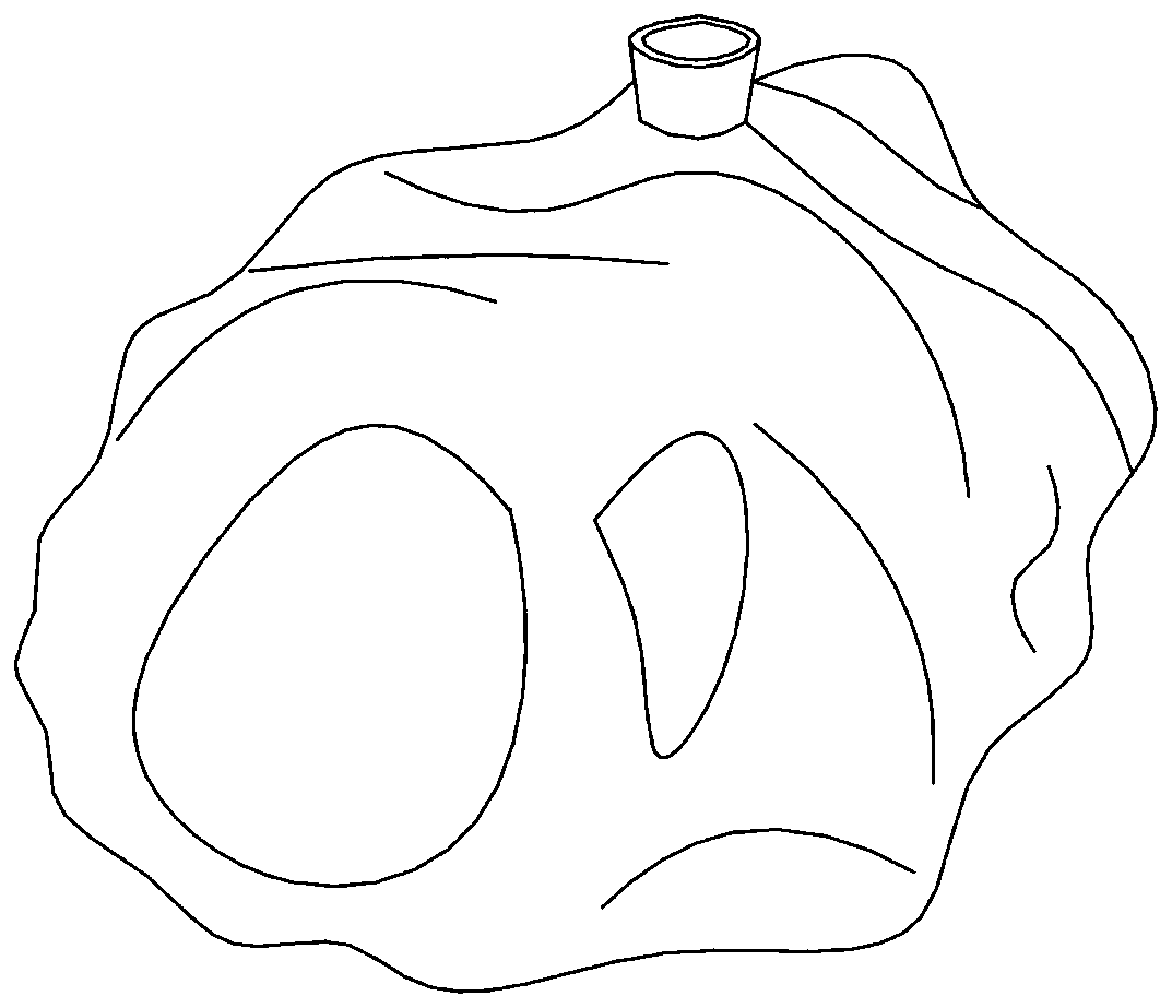
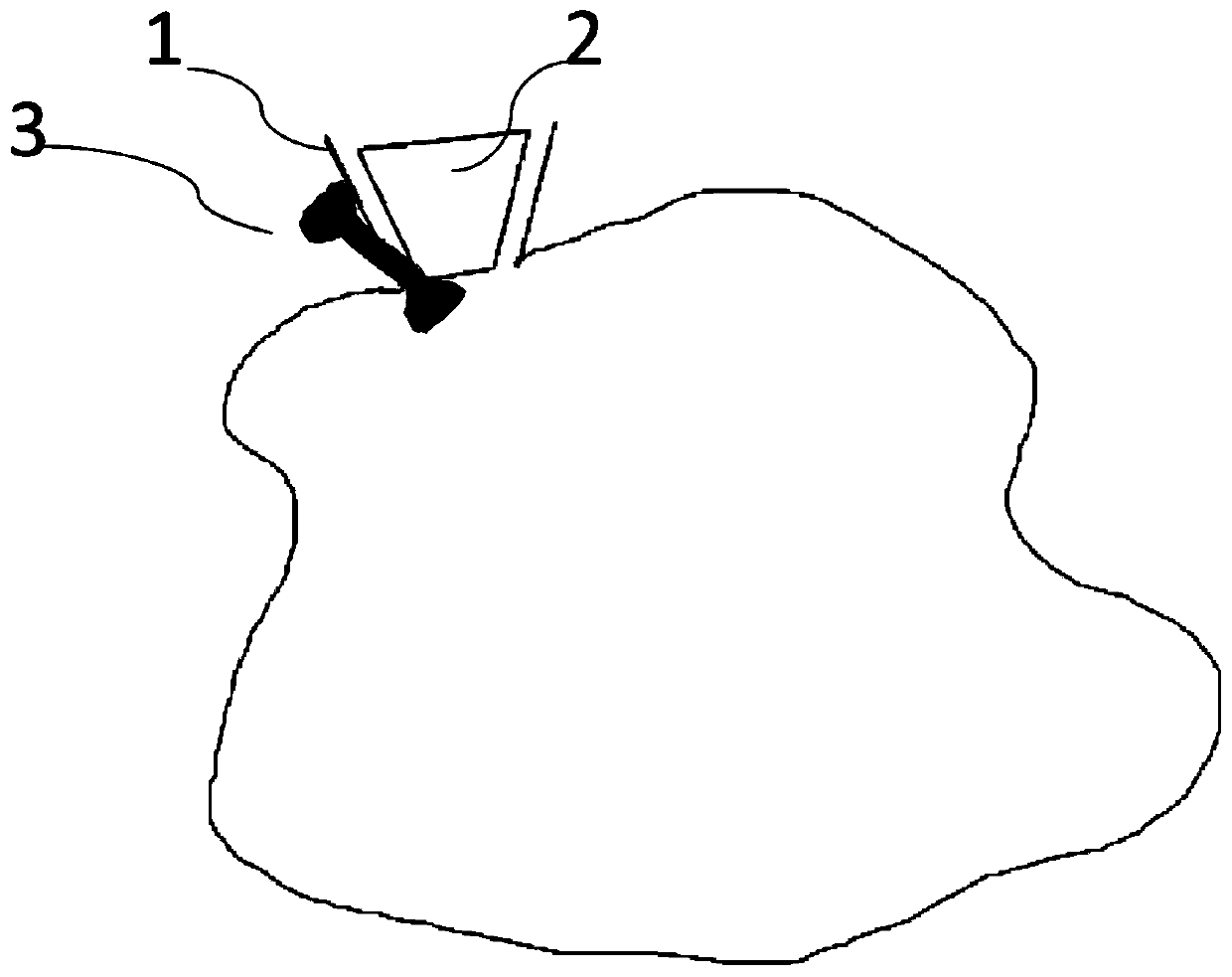
[0064] A simulated brain model, the simulated brain model includes a brain model with stretchable brain tissue and a simulated tumor device inside the brain model, the simulated tumor device is in contact with the stretchable brain tissue, and the simulated tumor device It includes a main body with a cavity made of soft material, an opening 1 arranged on the surface of the main body and an occluder 2 for blocking the opening.

[0065] Select a real tumor that needs to be simulated or displayed, and use DICOM format data to reconstruct a 3D visual model of the simulated tumor device, in which the shape and size are restored according to the image data of the selected simulated tumor. The simulated tumor device is designed to be hollow, and the simulated tumor device The wall thickness is 0.3mm. Opening 1 is used to empty the air in the simulated tumor device and then restore the simulated tumor device to the original design shape and size. The occluder is used to seal the simula...

Embodiment 2

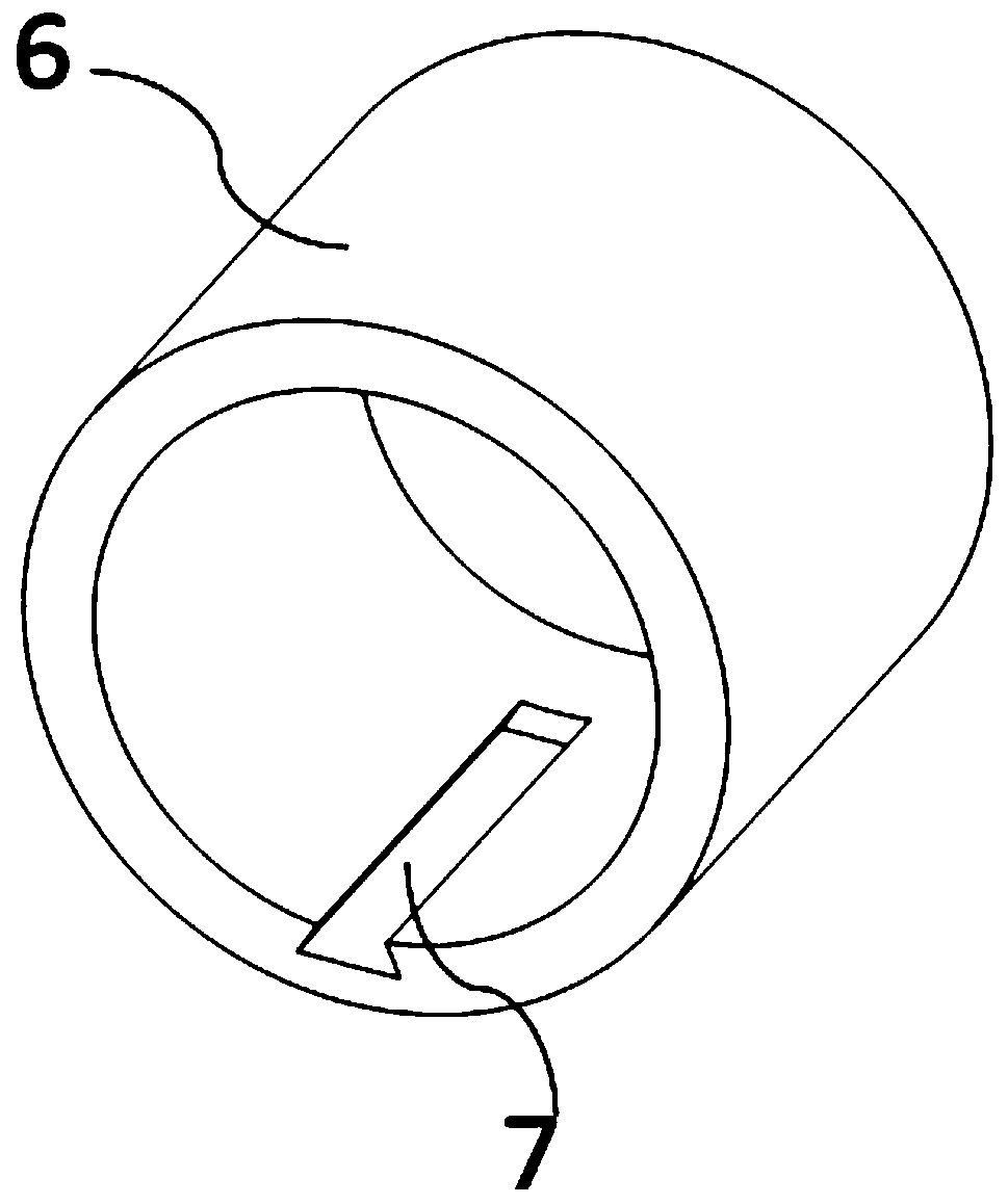
[0068] A simulated brain model, the simulated brain model includes a brain model with stretchable brain tissue and a simulated tumor device inside the brain model, the simulated tumor device is in contact with the stretchable brain tissue, and the simulated tumor device It includes a main body with a cavity made of soft material, an opening 1 on the surface of the main body and an occluder 2 for blocking the opening. Fill the tumor-simulating liquid to restore the original design shape and size of the tumor-simulating device.

[0069] Select a real tumor that needs to be simulated or displayed, and use DICOM format data to reconstruct a 3D visual model of the simulated tumor device. The size and shape are restored according to the image data of the selected simulated tumor. The simulated tumor device is designed to be hollow, and the simulated tumor device The wall thickness is 0.3mm, and there is also a horizontal bar 3 at the opening 1, which is like a dumbbell, and the hori...

Embodiment 3

[0075] A method for preparing a simulated brain model containing a simulated tumor device, comprising the steps of:

[0076] S1. Prepare a brain model containing stretchable brain tissue;

[0077] S2. According to the type of simulated tumor device, design and prepare the main body of the simulated tumor device with a cavity, the opening and the occluder on the surface of the main body, exhaust the air in the cavity of the main body of the simulated tumor device, and obtain a sheet-shaped simulated tumor device ;

[0078] S3. Make an opening on the stretchable brain tissue, place the sheet-shaped tumor-simulating device at the target position through the gap, and then fill the sheet-like tumor-simulating device with tumor-simulating liquid to restore the original design shape of the tumor-simulating device and The size, the opening is blocked with an occluder, and the brain tissue embedding simulated tumor device that can be pulled is completed;

[0079]S4. Filling the gaps ...


PUM

PropertyMeasurementUnit
Wall thicknessaaaaaaaaaa
Outer diameteraaaaaaaaaa
The inside diameter ofaaaaaaaaaa

Abstract

The invention discloses a simulated brain model and a preparation method thereof. The simulated brain model comprises a brain model body containing tractable brain tissue and a tumor simulating devicearranged inside the brain model body, wherein the tumor simulating device is in contact with the tractable brain tissue, and comprises a main body made of a soft material with a cavity, an opening formed in the surface of the main body and a stopper for blocking the opening. According to the simulated brain model, the elasticity and the touch feeling of real brain tissue are simulated to the greatest extent; the tumor simulating device is positioned in the brain model body and inflated and formed, so that the state that a real tumor strongly braces and extrudes the brain tissue is simulated to achieve the simulation effect; the tumor simulating device can be in any shape, variety, size and position, and is flexible and convenient; and after being used in a simulating tumor resection operation, the simulated brain model can be used for establishing tumor simulants of other parts or of different sizes and shapes, and the brain model containing a plurality of tumor simulating devices canalso be prepared to achieve the effect of flexible and recyclable use.

Description

technical field [0001] The invention relates to the technical field of clinical medicine, in particular to a simulated brain model and a preparation method thereof. Background technique [0002] With the development of neurosurgical equipment and technology, a large number of intracranial diseases can be solved by craniocerebral surgery. The cadaveric head model can provide preoperative practice conditions for craniocerebral surgery. However, the structure and position of the cranial brain will change after the cadaveric head is preserved. Increasingly, most neurosurgeons currently train surgically through computer simulations and autopsies. However, many craniocerebral diseases are specific, and the corresponding surgical or teaching models or cadavers are almost difficult to express realistically. In recent years, with the development of 3D printing technology, some human tissues and organs can be 3D printed and shaped, which has become an important method to assist doct...

Claims


Application Information

Patent Timeline
IPC IPC(8): B29C64/386B29C64/10B28B1/00B22F3/00G09B23/34B33Y50/00B33Y80/00B33Y10/00B22F10/00
CPCB29C64/386B29C64/10B28B1/001G09B23/34B33Y50/00B33Y80/00B33Y10/00B22F10/00B22F10/10Y02P10/25
Inventor 兰青
Owner THE SECOND HOSPITAL AFFILIATED TO SUZHOU UNIV
Why Eureka
Try Eureka