
Three-dimensional ultrasonic stereoscopic anatomical map generation method and device

A three-dimensional ultrasound and three-dimensional technology, which is applied in ultrasonic/sonic/infrasonic diagnosis, sonic diagnosis, infrasonic diagnosis, etc., can solve the problems affecting the accuracy of clinical diagnosis, cannot accurately determine the specific position of the section, etc., and achieve good results in clinical diagnosis.

Pending Publication Date: 2020-07-31
WUHAN ZHONGQI BIOLOGICAL MEDICAL ELECTRONICS
View PDF4 Cites 0 Cited by
  • Summary
  • Abstract
  • Description
  • Claims
  • Application Information

AI Technical Summary

Problems solved by technology

[0005] The purpose of the present invention is to overcome the above-mentioned technical deficiencies, provide a method and device for generating a three-dimensional ultrasonic stereoscopic anatomical map, and a computer storage medium, so as to solve the problem in the prior art that the section image and the three-dimensional view cannot be accurately judged in the three-dimensional anatomical space. Specific location, technical issues affecting the accuracy of clinical diagnosis

Method used


Image

  • Three-dimensional ultrasonic stereoscopic anatomical map generation method and device
  • Three-dimensional ultrasonic stereoscopic anatomical map generation method and device
  • Three-dimensional ultrasonic stereoscopic anatomical map generation method and device

Examples


Embodiment 1

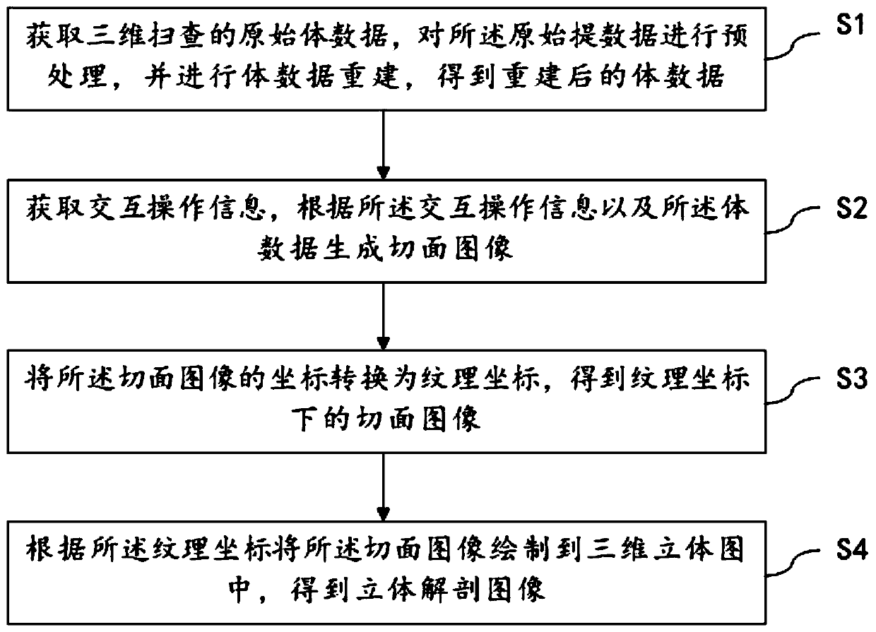
[0022] Such as figure 1 As shown, Embodiment 1 of the present invention provides a method for generating a three-dimensional ultrasonic stereoscopic anatomy map, comprising the following steps:

[0023] S1. Obtain the original volume data of the 3D scan, preprocess the original extracted data, and reconstruct the volume data to obtain the reconstructed volume data;

[0024] S2. Acquire interactive operation information, and generate a section image according to the interactive operation information and the volume data;

[0025] S3. Convert the coordinates of the section image into texture coordinates to obtain the section image under the texture coordinates;

[0026] S4. Draw the section image into a three-dimensional view according to the texture coordinates to obtain a three-dimensional anatomical map.

[0027] In this embodiment, the original volume data obtained by the three-dimensional scanning of the probe under the set scanning parameters is first obtained, and prepro...

Embodiment 2

[0061] Embodiment 2 of the present invention provides a device for generating a three-dimensional ultrasonic stereoscopic anatomy map, including a processor and a memory, and a computer program is stored in the memory. Method for generating ultrasound stereoscopic anatomy maps.

[0062] The three-dimensional ultrasonic stereoscopic anatomical map generation device provided by the embodiments of the present invention is used to realize the method for generating a three-dimensional ultrasonic stereoscopic anatomical map. Therefore, the technical effect of the three-dimensional ultrasonic stereoscopic anatomical map generation method is also possessed by the three-dimensional ultrasonic stereoscopic anatomical map generation device. I won't repeat them here.

Embodiment 3

[0064] Embodiment 3 of the present invention provides a computer storage medium on which a computer program is stored. When the computer program is executed by a processor, the method for generating a three-dimensional ultrasonic stereoscopic anatomy map provided in Embodiment 1 is implemented.

[0065] The computer storage medium provided by the embodiment of the present invention is used in the method for generating a three-dimensional ultrasonic stereoscopic anatomical map. Therefore, the technical effects of the method for generating a three-dimensional ultrasonic stereoscopic anatomical map are also provided by the computer storage medium, and will not be repeated here.


PUM


Abstract

The invention relates to the technical field of three-dimensional ultrasonic imaging, and discloses a three-dimensional ultrasonic stereoscopic anatomical map generation method. The method comprises the following steps of acquiring original volume data of three-dimensional scanning, preprocessing the original volume data, and reconstructing the volume data to obtain reconstructed volume data; acquiring interactive operation information, and generating a section image according to the interactive operation information and the volume data; converting the coordinates of the section image into texture coordinates to obtain a section image under the texture coordinates; and drawing the section image into a three-dimensional view according to the texture coordinates to obtain a stereoscopic anatomical map. According to the stereoscopic anatomical map generated by the method, the spatial position of the section image in the three-dimensional view is reflected, and a better basis is provided for accurate clinical diagnosis of an ultrasonic doctor.

Description

technical field [0001] The invention relates to the technical field of three-dimensional ultrasonic imaging, in particular to a method, device and computer storage medium for generating a three-dimensional ultrasonic stereoscopic anatomical map. Background technique [0002] Traditional ultrasound imaging equipment can only provide two-dimensional medical images. Ultrasound doctors can only estimate the size and shape of the lesion from multiple two-dimensional images based on experience, and use this to imagine the three-dimensional geometric relationship between the lesion and its surrounding tissue. The doctor's diagnosis posed difficulties. With the development of medical imaging technology, three-dimensional ultrasound imaging technology has become a mainstream ultrasound examination method in clinical practice. As an important part of the 3D ultrasound imaging system, 3D visualization technology can not only intuitively and vividly display the overall surface of the i...

Claims


Application Information

Patent Timeline
IPC IPC(8): A61B8/00
CPCA61B8/466A61B8/5253
Inventor 黄雄文刘王峰范兆龙张鹏鹏
Owner WUHAN ZHONGQI BIOLOGICAL MEDICAL ELECTRONICS
Learn More