An automatic medical image segmentation method based on multi-path attention fusion
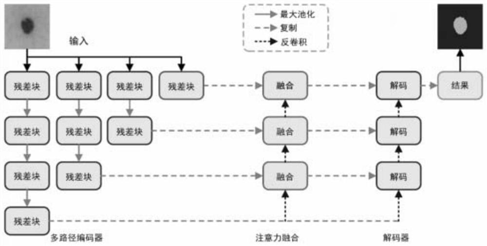
An automatic segmentation and medical image technology, applied in image analysis, neural learning methods, image enhancement, etc., can solve problems such as difficulty in saving spatial information, encoder loses spatial information, and affects segmentation results, so as to improve feature quality and increase image quality. Quantity, good accuracy effect
- Summary
- Abstract
- Description
- Claims
- Application Information
AI Technical Summary
Problems solved by technology
Method used
Image
Examples
Embodiment 1
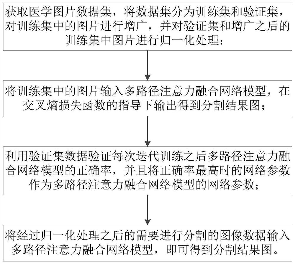
[0059] The pictures in the medical image data set are divided into a training set and a verification set. The training set is used to train the model, and the verification set is used to optimize the various indicators of the model. For medical image segmentation, it is not easy to obtain enough training samples. Therefore, the present invention Augment the pictures in the training set. The augmented operations include:
[0060] Rotate the pictures in the training set, the rotation angles include 10°, 20°, -10° and -20°, and save the rotated pictures;
[0061] Flip the pictures in the training set up and down and left and right, and save the flipped pictures;
[0062] Perform elastic transformation on the pictures in the training set, and save the pictures after elastic transformation;
[0063] Perform (20%, 80%) range scaling on the pictures in the training set, and save the scaled pictures;
[0064] Use the pictures in the training set and the pictures in the training set ...
Embodiment 2
[0083] Using the separation method in Example 1, in this implementation, Keras and Tensorflow open source deep learning libraries are used, NIVIDIA Geforce RTX-2080Ti GPU is used for training, Adam optimization algorithm model is used, and the learning rate is set to 0.0001; 2018ISIC skin is used Cancer lesion segmentation, LUNA lung CT dataset.
[0084] A data set in this example is provided by the 2018 Skin Cancer Lesion Segmentation Challenge, which contains a total of 2954 skin cancer lesion pictures, each with a size of 700×900 and a corresponding segmentation label map; using 1815 1 picture is used as a training set, 59 pictures are used as a verification set, and the remaining 520 pictures are used as a test set. In order to facilitate network training, the size of all pictures is adjusted to 256×256. The data in the test set is as follows: Figure 4 shown, where Figure 4 In the first row, the original image data, the second row is the label of the original image, the...
Embodiment 3
[0090] Using the separation method in Embodiment 1, different from Embodiment 2, this embodiment uses the LUNA dataset, which is provided by the 2017 Kaggle Lung Node Competition. Contains a total of 730 pictures and 730 corresponding segmentation label maps. The pixel size of each picture is 512×512. Use 70% of the pictures as the training set, 10% of the pictures as the verification set, and the remaining 20% of the data as a test set.
[0091] Due to the small amount of data, techniques such as rotation, flipping and elastic transformation are used to augment the training data set, so that the network can have good robustness and segmentation accuracy.
[0092] Four evaluation indicators are used, F1-score, Accuracy, Sensitivity and Specificity. The larger these four indexes are, the more accurate the segmentation effect is. As can be seen from Table 2, the experimental results in the LUNA data set show that compared with U-Net, R2-Unet, BCD-Net and U-Net++, the method of...
PUM
 Abstract
Description
Claims
Application Information
 - R&D
- Intellectual Property
- Life Sciences
- Materials
- Tech Scout
- Unparalleled Data Quality
- Higher Quality Content
- 60% Fewer Hallucinations
Browse by: Latest US Patents, China's latest patents, Technical Efficacy Thesaurus, Application Domain, Technology Topic, Popular Technical Reports.
© 2025 PatSnap. All rights reserved.Legal|Privacy policy|Modern Slavery Act Transparency Statement|Sitemap|About US| Contact US: help@patsnap.com


