Preparation method of tumor organoid
A technology of organoids and tumors, which is applied in the field of tumor organoid preparation, can solve the problems of difficult detection of tumor organoids, and achieve the effects of short culture time, good connection, and not easy to scatter
- Summary
- Abstract
- Description
- Claims
- Application Information
AI Technical Summary
Problems solved by technology
Method used
Image

Examples
Embodiment 1
[0037] Example 1: Preparation of tumor organoids
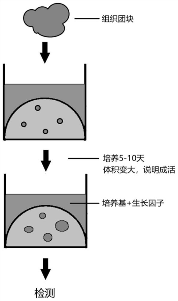
[0038] 1) Obtain a biopsy from a cancer or tumor patient.
[0039] 2) The biopsy tissue is soaked in tissue preservation solution to ensure tissue activity.
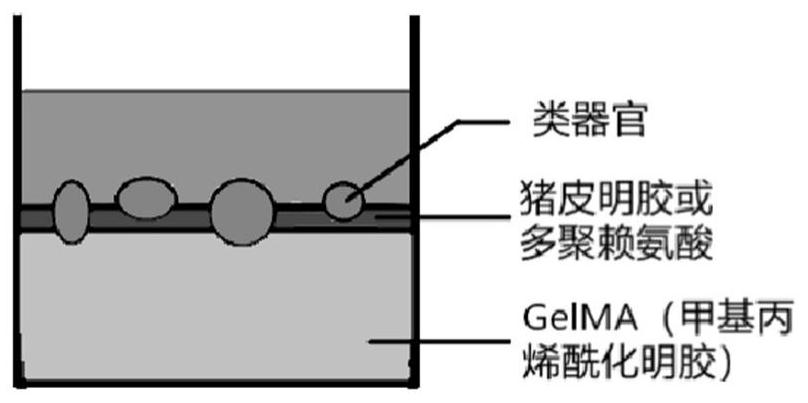
[0040] 3) Spread a layer of methacrylylated gelatin on a cell culture plate (96-well plate, 48-well plate, 24-well plate, 12-well plate, or 6-well plate), irradiate blue light to solidify the gel, and then spread A layer of pigskin gelatin is applied to adhere to the surface of the methacrylated gelatin.
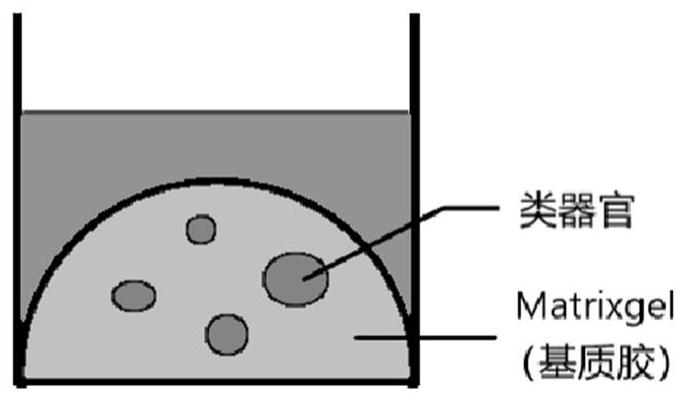
[0041] 4) Take the tissue out of the preservation solution, place it in a cell culture dish, add cell culture medium, chop the tissue in the cell culture medium, and the volume of small clumps is below 0.5mm×0.5mm.
[0042] 5) Mix the minced tissue-medium suspension and pass it through a sterile filter with a pore size of 100 um.
[0043] 6) The liquid passing through the filter is either discarded or used to cultivate primary cells, and the tissue mass intercepted ...
Embodiment 2
[0055] Embodiment 2: drug screening
[0056] For the testing of some new drugs, commonly used models in laboratories are PDX models (Patient-Derived tumor Xenograft) or CDX models (Cell-line-Derived tumor Xenograft). The PDX model is to extract the patient's primary cells and transplant them under the skin of nude mice or other immune-deficient mice to make the cells grow into tumors. After that, the mice were injected with the drug, and after several cycles of injection, the mass and volume of the tumor were measured to see if it was reduced compared to the control group. The problem with this is that although the experimental data count as in vivo experimental data, the growth environment in mice is very different from the growth environment in humans. For example, normal human neural stem cells cannot grow when transplanted into mice. Therefore, it is unknown whether the characteristics of patient tumor cells that can grow smoothly in mice have changed.
[0057] The grow...
Embodiment 3
[0059] Example 3: Drug Testing
[0060] With the patient's informed consent, the Shantou Cancer Hospital obtained the patient's recurrent cervical cancer tissue sample, put the tissue sample into the tissue preservation solution, and then obtained the tumor organoid according to the index method in Example 1. This tumor organoid was used for dosing experiments. The tested drugs included gemcitabine, paclitaxel, gemcitabine-romidepsin-cisplatin combination, C188-9, ivermectin-cisplatin combination, and BBI608-paclitaxel combination. Among them, C188-9 and BBI608 are new small molecular drugs, and the others are chemotherapy drugs commonly used in hospitals. Test results such as Figure 11 As shown, it can be seen from the figure that the tumor organoid is more sensitive to the combination of ivermectin-cisplatin and BBI608-paclitaxel, and the effective C188-9 in the tumor organoid test in Example 2 No obvious drug effect was shown.
PUM
 Abstract
Description
Claims
Application Information
 - R&D Engineer
- R&D Manager
- IP Professional
- Industry Leading Data Capabilities
- Powerful AI technology
- Patent DNA Extraction
Browse by: Latest US Patents, China's latest patents, Technical Efficacy Thesaurus, Application Domain, Technology Topic.
© 2024 PatSnap. All rights reserved.Legal|Privacy policy|Modern Slavery Act Transparency Statement|Sitemap