Cell image segmentation method and device and cell counting method

A technology for image segmentation and cell number, which is applied in image analysis, image data processing, image enhancement, etc., can solve the problems of lack of correction in estimation methods, large error in estimation results, and various overlapping cell shapes, etc., to achieve accurate cell statistics, The effect of removing mis-segmented regions

Active Publication Date: 2022-02-01
APPLITECH BIOLOGICAL TECH CO LTD
View PDF9 Cites 0 Cited by
  • Summary
  • Abstract
  • Description
  • Claims
  • Application Information

AI Technical Summary

Problems solved by technology

However, due to AO staining, living cells and dead cells will appear in clusters, the imaging background is complex, and the cell shapes overlap and vary. The general image segmentation method cannot identify and distinguish the number of cells in the clustered cells, so it can only be estimated by an algorithm. , but the existing estimation methods lack correction, and the estimation results have large errors

Method used


Image

  • Cell image segmentation method and device and cell counting method
  • Cell image segmentation method and device and cell counting method
  • Cell image segmentation method and device and cell counting method

Examples


Embodiment 1

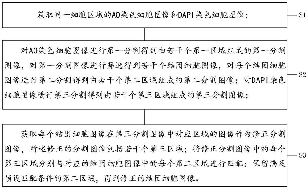
[0040] Such as figure 1 As shown, the present invention discloses a cell image segmentation method, which is applied to the segmentation and counting of stained cell images, and mainly includes the following steps:

[0041] Step S1, acquiring the AO-stained cell image and the DAPI-stained cell image of the same cell area;

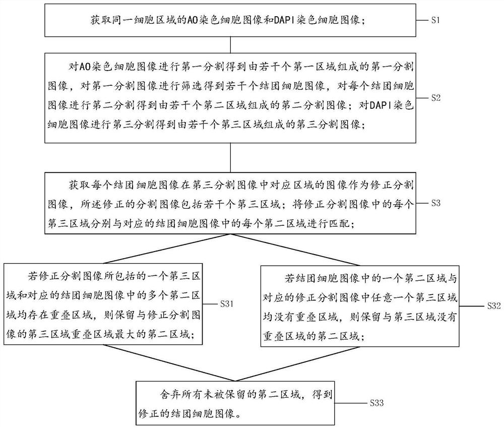
[0042] Step S2, performing the first segmentation on the AO-stained cell image to obtain a first segmented image composed of several first regions, screening the first segmented image to obtain several clustered cell images, and performing a second segmented image on each clustered cell image second segmentation to obtain a second segmented image composed of several second regions; performing a third segment on the DAPI-stained cell image to obtain a third segmented image composed of several third regions;

[0043] Step S3, acquiring the image of each clumped cell image corresponding to the third segmented image as the corrected segmented image, the correc...

Embodiment 2

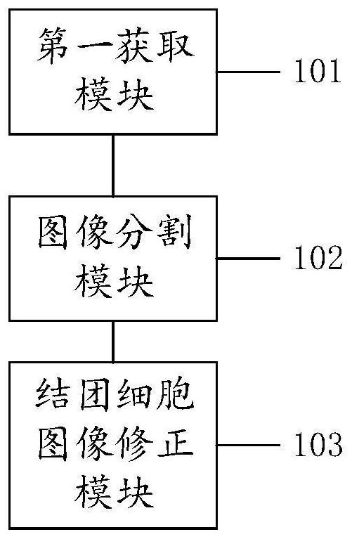
[0077] refer to image 3 , the present invention discloses a cell image segmentation device, comprising: a first acquisition module 101 , an image segmentation module 102 and an image correction module 103 of clumping cells.

[0078] The first acquiring module 101 is configured to acquire the AO stained cell image and the DAPI stained cell image of the same cell area;

[0079] The image segmentation module 102 is configured to first segment the AO stained cell image to obtain a first segmented image composed of several first regions, and to filter the first segmented image to obtain several clustered cell images, and for each Carrying out the second segmentation of the clumped cell image to obtain a second segmented image composed of several second regions; performing a third segment on the DAPI-stained cell image to obtain a third segmented image composed of several third regions;

[0080] The agglomerated cell image correction module 103 acquires an image of each agglomerat...

Embodiment 3

[0087]The present invention also discloses a computer-readable storage medium, the computer-readable storage medium includes a stored computer program, wherein, when the computer program is running, the device where the computer-readable storage medium is located is controlled to execute as in Embodiment 1. The described clumping cell image segmentation method.

[0088] Those of ordinary skill in the art can understand that realizing all or part of the processes in the above embodiments can be completed by instructing related hardware through a computer program. The computer program can be stored in a computer-readable storage medium. When the program is executed , may include the processes of the above-mentioned Embodiment 1. Wherein, the storage medium may be a magnetic disk, an optical disk, a read-only memory (Read-Only Memory, ROM), or a random access memory (Random Access Memory, RAM).


PUM


Abstract

The invention belongs to the technical field of cell image processing, and discloses a cell image segmentation method and device and a cell counting method, and the method comprises the steps: obtaining an AO stained cell image and a DAPI stained cell image of the same cell region; performing second segmentation on the clustered cell image in the AO staining cell image to obtain a second segmented image composed of second regions; performing third segmentation on the DAPI stained cell image to obtain a third segmented image composed of third regions; and obtaining a corrected segmented image, and matching each third region in the corrected segmented image with each second region in the corresponding clustered cell image to obtain a corrected clustered cell image. The method has the beneficial effects that the AO image is matched through the DAPI image, and the second region obtained through algorithm segmentation is corrected, so that a mistaken segmentation region caused by an algorithm is effectively eliminated, and a more accurate clustering cell image segmentation result can be obtained.

Description

technical field [0001] The invention relates to the technical field of cell image processing, in particular to a cell image segmentation method, device and cell counting method. Background technique [0002] At present, the use of cytological computer-aided diagnostic counting can effectively improve the efficiency and accuracy of cell counting by doctors or scientific researchers. When performing cell statistics, the existing counts generally use AO stained cell images for cell counting. AO staining can stain live cells and dead cells at the same time, and DAPI staining can only stain dead cells. However, due to AO staining, living cells and dead cells will appear in clusters, the imaging background is complex, and the cell shapes overlap and vary. The general image segmentation method cannot identify and distinguish the number of cells in the clustered cells, so it can only be estimated by an algorithm. , but the existing estimation methods lack correction, and the estim...

Claims


Application Information

Patent Timeline
Patent Type & Authority Applications(China)
IPC IPC(8): G06T7/11G06T7/174G06V10/75G06K9/62
CPCG06T7/11G06T7/174G06T2207/30024G06T2207/30242G06F18/22
Inventor 周文静张欣史振志
Owner APPLITECH BIOLOGICAL TECH CO LTD
Why Eureka
Try Eureka