Sub-pixel temporal skew correction for positron emission tomography (PET)

A sub-pixel and pixel technology, which is used in the field of gamma ray detectors and medical imaging equipment, can solve the problems of inability to realize clock distribution and deterioration of system performance.

Pending Publication Date: 2022-07-22
KONINKLJIJKE PHILIPS NV
View PDF1 Cites 0 Cited by
  • Summary
  • Abstract
  • Description
  • Claims
  • Application Information

AI Technical Summary

Problems solved by technology

Wave impedance and cable length cause time delays, and clock distribution along individual lanes cannot be achieved without additional buffers with corresponding individual time delays
[0005] Even though TOF PET can reduce the acquisition time and increase the effective sensitivity of PET acquisition by calculating the time difference between two coincident photons, for a system with a coincidence timing resolution of 200ps (FWHM), an uncorrected time skew of 20ps to 40ps can degrade system performance

Method used


Image

  • Sub-pixel temporal skew correction for positron emission tomography (PET)
  • Sub-pixel temporal skew correction for positron emission tomography (PET)
  • Sub-pixel temporal skew correction for positron emission tomography (PET)

Examples


Embodiment Construction

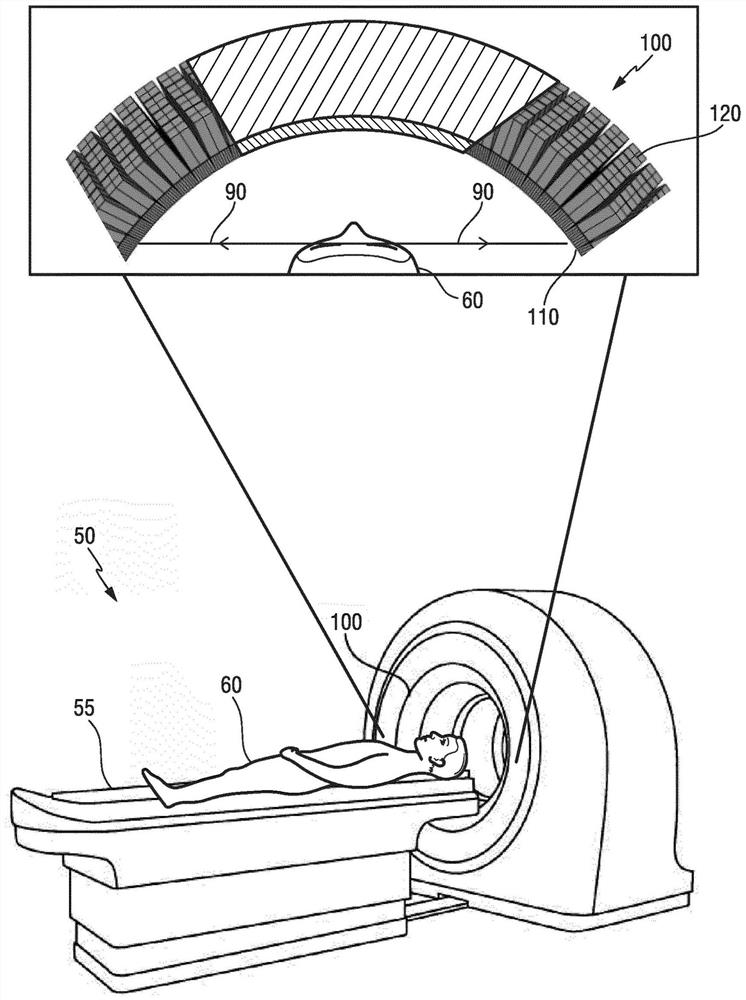
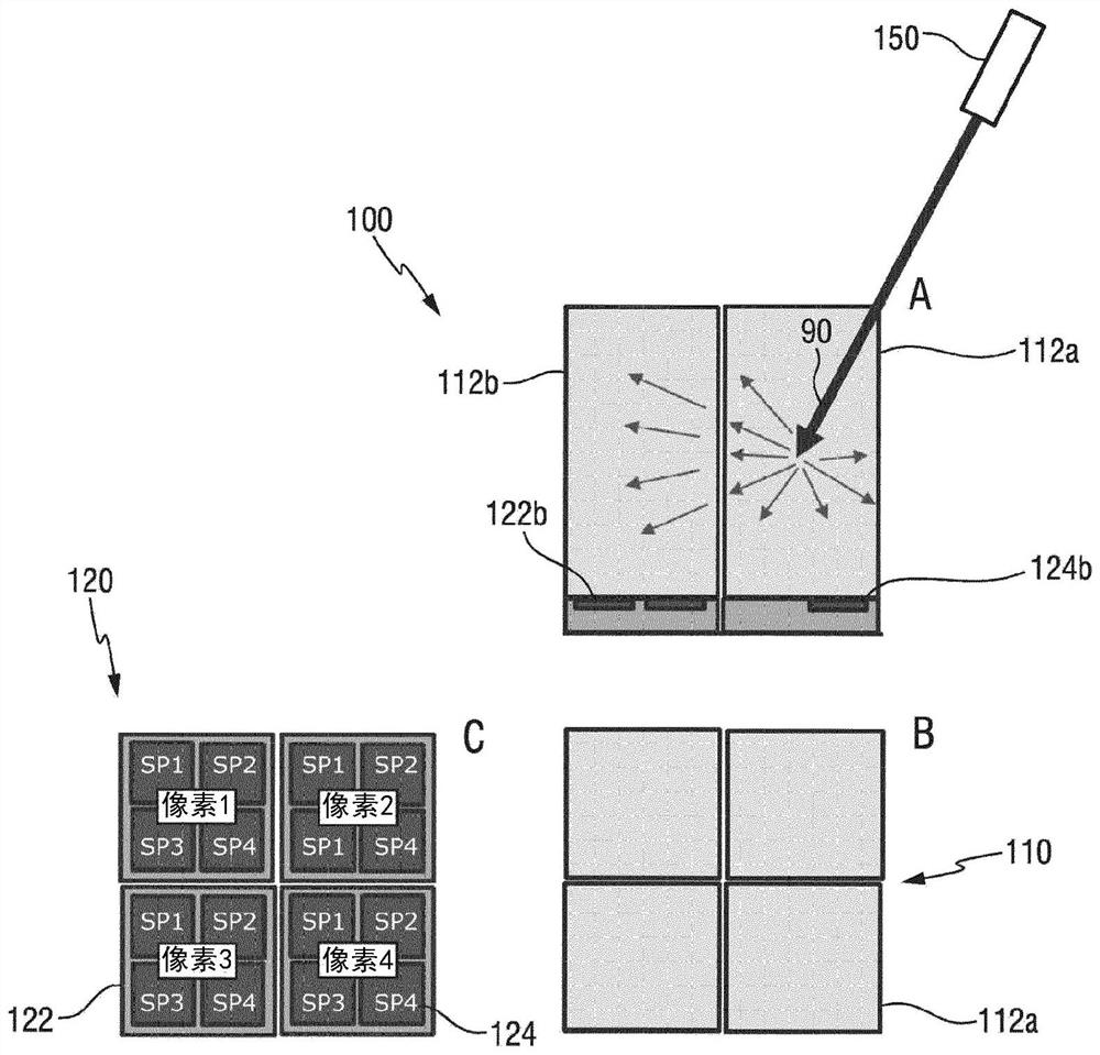
[0076] figure 1 A schematic diagram of a medical imaging device 50 according to the present invention is shown. The medical imaging device 50 may be, for example, a PET device, a PET / CT device, a PET / MR device or a SPECT / PET / CT device. The illustrated medical imaging device 50 includes a gamma-ray detector 100 that is preferably calibrated by using a calibration method or calibration module according to the present invention.

[0077] The illustrated medical imaging device 50 also includes a tunable patient support 55 for supporting a patient 60 undergoing treatment by means of the medical imaging device 50 . PET devices typically detect particles, particularly gamma rays emitted within the patient 60 to be imaged. For example, the patient 60 may be administered a radioactive tracer substance, and a gamma ray detector 100 calibrated in accordance with the present invention may be used to determine the spatial location of the tracer substance in the patient 60 . Therefore, P...


PUM


Abstract

The present invention relates to a calibration method for a gamma ray detector (100), the gamma ray detector comprising: a pixelated scintillator array (110) for emitting scintillation photons at a photon conversion location (94) in response to incident gamma rays (90); and a pixelated photodetector array (120) for determining a spatial intensity distribution of the scintillation photons. The present invention is based on the concept of using the concept of optical light sharing of scintillation photons (in an element (i.e., in an optical field). ) of the scintillator array (110), one scintillator pixel (112) of the scintillator array (110), and distributed over a plurality of photodetector pixels (122) of the pixelated photodetector array (120), allow for obtaining an estimate for a temporal skew between adjacent photodetector pixels (122). The invention also relates to a calibration module (200) for a gamma ray detector (100) comprising a recorder (210) and a processing module (220) to perform the functions of the above method. In addition, the invention also relates to a gamma ray detector (100) and a medical imaging device (50) comprising the gamma ray detector (100).

Description

technical field [0001] The present invention relates to a calibration method for a gamma ray detector, a corresponding calibration module for a gamma ray detector, a gamma ray detector and a medical imaging device. Background technique [0002] Positron emission tomography (PET) is a commonly used medical imaging technique. The technique is based on the detection of gamma-ray photons emitted after administration of a radiotracer substance to a patient (eg, by injecting the radiotracer substance into a peripheral vein). The nuclei of the radioisotope of the radiotracer substance emit positrons (positively charged electrons) that collide with electrons in the patient's tissue. This process converts mass into energy in the form of two emitted gamma-ray photons. The two gamma-ray photons are directed in opposite directions and are detected by a radiation detector surrounding the imaging object (patient) as substantially simultaneous radiation detection events that define a lin...

Claims


Application Information

Patent Timeline
Patent Type & Authority Applications(China)
IPC IPC(8): G01T1/164G01T7/00
CPCG01T7/005G01T1/1647G01T1/1642G01T1/1648
Inventor T·佐尔夫T·弗拉奇
Owner KONINKLJIJKE PHILIPS NV
Why Eureka
Try Eureka